Stuart Baker, Ricardo Núñez Miguel, Daniel Thomas, Michael Powell, Jadwiga Furmaniak, Bernard Rees Smith
{"title":"人甲状腺过氧化物酶(TPO)与TPO抗体复合物的低温电镜结构。","authors":"Stuart Baker, Ricardo Núñez Miguel, Daniel Thomas, Michael Powell, Jadwiga Furmaniak, Bernard Rees Smith","doi":"10.1530/JME-22-0149","DOIUrl":null,"url":null,"abstract":"Determination of the structure of the extracellular domain of human thyroid peroxidase (hTPO) by cryo-electron microscopy (cryo-EM) is described. TPO, purified to homogeneity was complexed with the hTPO monoclonal autoantibody 2G4 Fab and also with a mouse monoclonal TPO antibody 4F5 Fab (which competes with autoantibody binding to TPO). Both complexes were analysed by cryo-EM. The two structures (global resolution 3.92 and 3.4 Å for the 2G4 complex and 4F5 complex, respectively) show TPO as a monomer with four domains; the N-terminal domain, the peroxidase domain (POD), the complement control protein (CCP)-like domain and the epidermal growth factor-like domain which are all visible in the structures. The relative positions of the domains are fixed with a disulphide bond between cysteine residues Cys146 in the POD and Cys756 in the CCP domain preventing significant flexibility of the molecule. The entrance to the enzyme active site, the haem group and the calcium binding site are clearly visible on the opposite side of the TPO molecule from the 2G4 and 4F5 binding sites. Extensive interactions are seen between TPO and the two antibodies which both bind to distinct epitopes on the POD domain, including some residues in the immunodominant region B mainly via different residues. However, the epitopes of the two antibodies contain three shared TPO residues. This is the first high-resolution structure of TPO to be reported and it should help guide the development of new inhibitors of TPO enzyme activity for therapeutic applications.","PeriodicalId":16570,"journal":{"name":"Journal of molecular endocrinology","volume":"70 3","pages":""},"PeriodicalIF":3.6000,"publicationDate":"2023-04-01","publicationTypes":"Journal Article","fieldsOfStudy":null,"isOpenAccess":false,"openAccessPdf":"https://www.ncbi.nlm.nih.gov/pmc/articles/PMC9986399/pdf/","citationCount":"1","resultStr":"{\"title\":\"Cryo-electron microscopy structures of human thyroid peroxidase (TPO) in complex with TPO antibodies.\",\"authors\":\"Stuart Baker, Ricardo Núñez Miguel, Daniel Thomas, Michael Powell, Jadwiga Furmaniak, Bernard Rees Smith\",\"doi\":\"10.1530/JME-22-0149\",\"DOIUrl\":null,\"url\":null,\"abstract\":\"Determination of the structure of the extracellular domain of human thyroid peroxidase (hTPO) by cryo-electron microscopy (cryo-EM) is described. TPO, purified to homogeneity was complexed with the hTPO monoclonal autoantibody 2G4 Fab and also with a mouse monoclonal TPO antibody 4F5 Fab (which competes with autoantibody binding to TPO). Both complexes were analysed by cryo-EM. The two structures (global resolution 3.92 and 3.4 Å for the 2G4 complex and 4F5 complex, respectively) show TPO as a monomer with four domains; the N-terminal domain, the peroxidase domain (POD), the complement control protein (CCP)-like domain and the epidermal growth factor-like domain which are all visible in the structures. The relative positions of the domains are fixed with a disulphide bond between cysteine residues Cys146 in the POD and Cys756 in the CCP domain preventing significant flexibility of the molecule. The entrance to the enzyme active site, the haem group and the calcium binding site are clearly visible on the opposite side of the TPO molecule from the 2G4 and 4F5 binding sites. Extensive interactions are seen between TPO and the two antibodies which both bind to distinct epitopes on the POD domain, including some residues in the immunodominant region B mainly via different residues. However, the epitopes of the two antibodies contain three shared TPO residues. This is the first high-resolution structure of TPO to be reported and it should help guide the development of new inhibitors of TPO enzyme activity for therapeutic applications.\",\"PeriodicalId\":16570,\"journal\":{\"name\":\"Journal of molecular endocrinology\",\"volume\":\"70 3\",\"pages\":\"\"},\"PeriodicalIF\":3.6000,\"publicationDate\":\"2023-04-01\",\"publicationTypes\":\"Journal Article\",\"fieldsOfStudy\":null,\"isOpenAccess\":false,\"openAccessPdf\":\"https://www.ncbi.nlm.nih.gov/pmc/articles/PMC9986399/pdf/\",\"citationCount\":\"1\",\"resultStr\":null,\"platform\":\"Semanticscholar\",\"paperid\":null,\"PeriodicalName\":\"Journal of molecular endocrinology\",\"FirstCategoryId\":\"3\",\"ListUrlMain\":\"https://doi.org/10.1530/JME-22-0149\",\"RegionNum\":4,\"RegionCategory\":\"医学\",\"ArticlePicture\":[],\"TitleCN\":null,\"AbstractTextCN\":null,\"PMCID\":null,\"EPubDate\":\"\",\"PubModel\":\"\",\"JCR\":\"Q2\",\"JCRName\":\"ENDOCRINOLOGY & METABOLISM\",\"Score\":null,\"Total\":0}","platform":"Semanticscholar","paperid":null,"PeriodicalName":"Journal of molecular endocrinology","FirstCategoryId":"3","ListUrlMain":"https://doi.org/10.1530/JME-22-0149","RegionNum":4,"RegionCategory":"医学","ArticlePicture":[],"TitleCN":null,"AbstractTextCN":null,"PMCID":null,"EPubDate":"","PubModel":"","JCR":"Q2","JCRName":"ENDOCRINOLOGY & METABOLISM","Score":null,"Total":0}
Cryo-electron microscopy structures of human thyroid peroxidase (TPO) in complex with TPO antibodies.
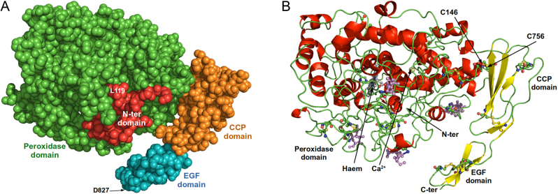
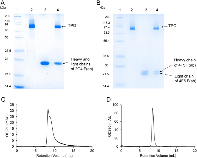
Determination of the structure of the extracellular domain of human thyroid peroxidase (hTPO) by cryo-electron microscopy (cryo-EM) is described. TPO, purified to homogeneity was complexed with the hTPO monoclonal autoantibody 2G4 Fab and also with a mouse monoclonal TPO antibody 4F5 Fab (which competes with autoantibody binding to TPO). Both complexes were analysed by cryo-EM. The two structures (global resolution 3.92 and 3.4 Å for the 2G4 complex and 4F5 complex, respectively) show TPO as a monomer with four domains; the N-terminal domain, the peroxidase domain (POD), the complement control protein (CCP)-like domain and the epidermal growth factor-like domain which are all visible in the structures. The relative positions of the domains are fixed with a disulphide bond between cysteine residues Cys146 in the POD and Cys756 in the CCP domain preventing significant flexibility of the molecule. The entrance to the enzyme active site, the haem group and the calcium binding site are clearly visible on the opposite side of the TPO molecule from the 2G4 and 4F5 binding sites. Extensive interactions are seen between TPO and the two antibodies which both bind to distinct epitopes on the POD domain, including some residues in the immunodominant region B mainly via different residues. However, the epitopes of the two antibodies contain three shared TPO residues. This is the first high-resolution structure of TPO to be reported and it should help guide the development of new inhibitors of TPO enzyme activity for therapeutic applications.
期刊介绍:
The Journal of Molecular Endocrinology is an official journal of the Society for Endocrinology and is endorsed by the European Society of Endocrinology and the Endocrine Society of Australia.
Journal of Molecular Endocrinology is a leading global journal that publishes original research articles and reviews. The journal focuses on molecular and cellular mechanisms in endocrinology, including: gene regulation, cell biology, signalling, mutations, transgenics, hormone-dependant cancers, nuclear receptors, and omics. Basic and pathophysiological studies at the molecule and cell level are considered, as well as human sample studies where this is the experimental model of choice. Technique studies including CRISPR or gene editing are also encouraged.


 分享
分享
 求助内容:
求助内容: 应助结果提醒方式:
 扫码关注我们
