Hajo Findeisen, Christina Westhoff, Corinna Trenker, Christian Görg, Johannes Krönig, Ehsan Safai Zadeh
{"title":"超声造影诊断肺囊性包虫病1例。","authors":"Hajo Findeisen, Christina Westhoff, Corinna Trenker, Christian Görg, Johannes Krönig, Ehsan Safai Zadeh","doi":"10.15557/JoU.2023.0008","DOIUrl":null,"url":null,"abstract":"<p><strong>Aim of the study: </strong>Pulmonary cystic echinococcosis is a parasitic infection transmitted by dogs and occurring in livestock-raising areas. It is included among the neglected tropical diseases, according to the World Health Organization. Imaging plays a pivotal role in the diagnosis of this disease. While cross-sectional imaging modalities such as computed tomography and magnetic resonance imaging are preferred, lung ultrasound may be another feasible technique.</p><p><strong>Case description: </strong>We report a case of pulmonary cystic echinococcosis in a 26-year-old woman who was examined by contrast-enhanced ultrasound, which showed marked annular enhancement around the hydatid cyst, mimicking a superinfected cyst.</p><p><strong>Conclusions: </strong>Contrast-enhanced ultrasound examination in pulmonary cystic echinococcosis should be studied in a larger population to determine the value of additional contrast administration. In the present case report, no superinfected echinococcal cyst was seen despite marked annular contrast enhancement.</p>","PeriodicalId":45612,"journal":{"name":"Journal of Ultrasonography","volume":"23 92","pages":"39-42"},"PeriodicalIF":1.5000,"publicationDate":"2023-01-01","publicationTypes":"Journal Article","fieldsOfStudy":null,"isOpenAccess":false,"openAccessPdf":"https://ftp.ncbi.nlm.nih.gov/pub/pmc/oa_pdf/9d/04/jou-23-039.PMC9985186.pdf","citationCount":"0","resultStr":"{\"title\":\"Pulmonary Cystic Echinococcosis in Contrast-enhanced Ultrasound - A Case Report.\",\"authors\":\"Hajo Findeisen, Christina Westhoff, Corinna Trenker, Christian Görg, Johannes Krönig, Ehsan Safai Zadeh\",\"doi\":\"10.15557/JoU.2023.0008\",\"DOIUrl\":null,\"url\":null,\"abstract\":\"<p><strong>Aim of the study: </strong>Pulmonary cystic echinococcosis is a parasitic infection transmitted by dogs and occurring in livestock-raising areas. It is included among the neglected tropical diseases, according to the World Health Organization. Imaging plays a pivotal role in the diagnosis of this disease. While cross-sectional imaging modalities such as computed tomography and magnetic resonance imaging are preferred, lung ultrasound may be another feasible technique.</p><p><strong>Case description: </strong>We report a case of pulmonary cystic echinococcosis in a 26-year-old woman who was examined by contrast-enhanced ultrasound, which showed marked annular enhancement around the hydatid cyst, mimicking a superinfected cyst.</p><p><strong>Conclusions: </strong>Contrast-enhanced ultrasound examination in pulmonary cystic echinococcosis should be studied in a larger population to determine the value of additional contrast administration. In the present case report, no superinfected echinococcal cyst was seen despite marked annular contrast enhancement.</p>\",\"PeriodicalId\":45612,\"journal\":{\"name\":\"Journal of Ultrasonography\",\"volume\":\"23 92\",\"pages\":\"39-42\"},\"PeriodicalIF\":1.5000,\"publicationDate\":\"2023-01-01\",\"publicationTypes\":\"Journal Article\",\"fieldsOfStudy\":null,\"isOpenAccess\":false,\"openAccessPdf\":\"https://ftp.ncbi.nlm.nih.gov/pub/pmc/oa_pdf/9d/04/jou-23-039.PMC9985186.pdf\",\"citationCount\":\"0\",\"resultStr\":null,\"platform\":\"Semanticscholar\",\"paperid\":null,\"PeriodicalName\":\"Journal of Ultrasonography\",\"FirstCategoryId\":\"1085\",\"ListUrlMain\":\"https://doi.org/10.15557/JoU.2023.0008\",\"RegionNum\":0,\"RegionCategory\":null,\"ArticlePicture\":[],\"TitleCN\":null,\"AbstractTextCN\":null,\"PMCID\":null,\"EPubDate\":\"\",\"PubModel\":\"\",\"JCR\":\"Q3\",\"JCRName\":\"RADIOLOGY, NUCLEAR MEDICINE & MEDICAL IMAGING\",\"Score\":null,\"Total\":0}","platform":"Semanticscholar","paperid":null,"PeriodicalName":"Journal of Ultrasonography","FirstCategoryId":"1085","ListUrlMain":"https://doi.org/10.15557/JoU.2023.0008","RegionNum":0,"RegionCategory":null,"ArticlePicture":[],"TitleCN":null,"AbstractTextCN":null,"PMCID":null,"EPubDate":"","PubModel":"","JCR":"Q3","JCRName":"RADIOLOGY, NUCLEAR MEDICINE & MEDICAL IMAGING","Score":null,"Total":0}
Pulmonary Cystic Echinococcosis in Contrast-enhanced Ultrasound - A Case Report.
Aim of the study: Pulmonary cystic echinococcosis is a parasitic infection transmitted by dogs and occurring in livestock-raising areas. It is included among the neglected tropical diseases, according to the World Health Organization. Imaging plays a pivotal role in the diagnosis of this disease. While cross-sectional imaging modalities such as computed tomography and magnetic resonance imaging are preferred, lung ultrasound may be another feasible technique.
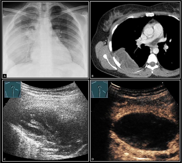
Case description: We report a case of pulmonary cystic echinococcosis in a 26-year-old woman who was examined by contrast-enhanced ultrasound, which showed marked annular enhancement around the hydatid cyst, mimicking a superinfected cyst.
Conclusions: Contrast-enhanced ultrasound examination in pulmonary cystic echinococcosis should be studied in a larger population to determine the value of additional contrast administration. In the present case report, no superinfected echinococcal cyst was seen despite marked annular contrast enhancement.


 分享
分享
 求助内容:
求助内容: 应助结果提醒方式:
 扫码关注我们
