Mustafa Çalışkan, Ömer Faruk Baycan, Fatma Betül Çelik, Tolga Sinan Güvenç, Adem Atıcı, Yasemin Çağ, Oğuz Konal, Tuğçe İrgi, Ümmühan Zeynep Bilgili, Mehmet Ali Ağırbaşlı
{"title":"冠状动脉微血管功能障碍在COVID-19感染住院患者中很常见","authors":"Mustafa Çalışkan, Ömer Faruk Baycan, Fatma Betül Çelik, Tolga Sinan Güvenç, Adem Atıcı, Yasemin Çağ, Oğuz Konal, Tuğçe İrgi, Ümmühan Zeynep Bilgili, Mehmet Ali Ağırbaşlı","doi":"10.1111/micc.12757","DOIUrl":null,"url":null,"abstract":"<div>\n \n \n <section>\n \n <h3> Background and Aims</h3>\n \n <p>Microvascular disease is considered as one of the main drivers of morbidity and mortality in severe COVID-19, and microvascular dysfunction has been demonstrated in the subcutaneous and sublingual tissues in COVID-19 patients. The presence of coronary microvascular dysfunction (CMD) has also been hypothesized, but direct evidence demonstrating CMD in COVID-19 patients is missing. In the present study, we aimed to investigate CMD in patients hospitalized with COVID-19, and to understand whether there is a relationship between biomarkers of myocardial injury, myocardial strain and inflammation and CMD.</p>\n </section>\n \n <section>\n \n <h3> Methods</h3>\n \n <p>39 patients that were hospitalized with COVID-19 and 40 control subjects were included to the present study. Biomarkers for myocardial injury, myocardial strain, inflammation, and fibrin turnover were obtained at admission. A comprehensive echocardiographic examination, including measurement of coronary flow velocity reserve (CFVR), was done after the patient was stabilized.</p>\n </section>\n \n <section>\n \n <h3> Results</h3>\n \n <p>Patients with COVID-19 infection had a significantly lower hyperemic coronary flow velocity, resulting in a significantly lower CFVR (2.0 ± 0.3 vs. 2.4 ± 0.5, <i>p</i> < .001). Patients with severe COVID-19 had a lower CFVR compared to those with moderate COVID-19 (1.8 ± 0.2 vs. 2.2 ± 0.2, <i>p</i> < .001) driven by a trend toward higher basal flow velocity. CFVR correlated with troponin (<i>p</i> = .003, <i>r</i>: −.470), B-type natriuretic peptide (<i>p</i> < .001, <i>r</i>: −.580), C-reactive protein (<i>p</i> < .001, <i>r</i>: −.369), interleukin-6 (<i>p</i> < .001, <i>r</i>: −.597), and d-dimer (<i>p</i> < .001, <i>r</i>: −.561), with the three latter biomarkers having the highest areas-under-curve for predicting CMD.</p>\n </section>\n \n <section>\n \n <h3> Conclusions</h3>\n \n <p>Coronary microvascular dysfunction is common in patients with COVID-19 and is related to the severity of the infection. CMD may also explain the “cryptic” myocardial injury seen in patients with severe COVID-19 infection.</p>\n </section>\n </div>","PeriodicalId":18459,"journal":{"name":"Microcirculation","volume":"29 4-5","pages":""},"PeriodicalIF":2.0000,"publicationDate":"2022-04-18","publicationTypes":"Journal Article","fieldsOfStudy":null,"isOpenAccess":false,"openAccessPdf":"https://onlinelibrary.wiley.com/doi/epdf/10.1111/micc.12757","citationCount":"7","resultStr":"{\"title\":\"Coronary microvascular dysfunction is common in patients hospitalized with COVID-19 infection\",\"authors\":\"Mustafa Çalışkan, Ömer Faruk Baycan, Fatma Betül Çelik, Tolga Sinan Güvenç, Adem Atıcı, Yasemin Çağ, Oğuz Konal, Tuğçe İrgi, Ümmühan Zeynep Bilgili, Mehmet Ali Ağırbaşlı\",\"doi\":\"10.1111/micc.12757\",\"DOIUrl\":null,\"url\":null,\"abstract\":\"<div>\\n \\n \\n <section>\\n \\n <h3> Background and Aims</h3>\\n \\n <p>Microvascular disease is considered as one of the main drivers of morbidity and mortality in severe COVID-19, and microvascular dysfunction has been demonstrated in the subcutaneous and sublingual tissues in COVID-19 patients. The presence of coronary microvascular dysfunction (CMD) has also been hypothesized, but direct evidence demonstrating CMD in COVID-19 patients is missing. In the present study, we aimed to investigate CMD in patients hospitalized with COVID-19, and to understand whether there is a relationship between biomarkers of myocardial injury, myocardial strain and inflammation and CMD.</p>\\n </section>\\n \\n <section>\\n \\n <h3> Methods</h3>\\n \\n <p>39 patients that were hospitalized with COVID-19 and 40 control subjects were included to the present study. Biomarkers for myocardial injury, myocardial strain, inflammation, and fibrin turnover were obtained at admission. A comprehensive echocardiographic examination, including measurement of coronary flow velocity reserve (CFVR), was done after the patient was stabilized.</p>\\n </section>\\n \\n <section>\\n \\n <h3> Results</h3>\\n \\n <p>Patients with COVID-19 infection had a significantly lower hyperemic coronary flow velocity, resulting in a significantly lower CFVR (2.0 ± 0.3 vs. 2.4 ± 0.5, <i>p</i> < .001). Patients with severe COVID-19 had a lower CFVR compared to those with moderate COVID-19 (1.8 ± 0.2 vs. 2.2 ± 0.2, <i>p</i> < .001) driven by a trend toward higher basal flow velocity. CFVR correlated with troponin (<i>p</i> = .003, <i>r</i>: −.470), B-type natriuretic peptide (<i>p</i> < .001, <i>r</i>: −.580), C-reactive protein (<i>p</i> < .001, <i>r</i>: −.369), interleukin-6 (<i>p</i> < .001, <i>r</i>: −.597), and d-dimer (<i>p</i> < .001, <i>r</i>: −.561), with the three latter biomarkers having the highest areas-under-curve for predicting CMD.</p>\\n </section>\\n \\n <section>\\n \\n <h3> Conclusions</h3>\\n \\n <p>Coronary microvascular dysfunction is common in patients with COVID-19 and is related to the severity of the infection. CMD may also explain the “cryptic” myocardial injury seen in patients with severe COVID-19 infection.</p>\\n </section>\\n </div>\",\"PeriodicalId\":18459,\"journal\":{\"name\":\"Microcirculation\",\"volume\":\"29 4-5\",\"pages\":\"\"},\"PeriodicalIF\":2.0000,\"publicationDate\":\"2022-04-18\",\"publicationTypes\":\"Journal Article\",\"fieldsOfStudy\":null,\"isOpenAccess\":false,\"openAccessPdf\":\"https://onlinelibrary.wiley.com/doi/epdf/10.1111/micc.12757\",\"citationCount\":\"7\",\"resultStr\":null,\"platform\":\"Semanticscholar\",\"paperid\":null,\"PeriodicalName\":\"Microcirculation\",\"FirstCategoryId\":\"3\",\"ListUrlMain\":\"https://onlinelibrary.wiley.com/doi/10.1111/micc.12757\",\"RegionNum\":4,\"RegionCategory\":\"医学\",\"ArticlePicture\":[],\"TitleCN\":null,\"AbstractTextCN\":null,\"PMCID\":null,\"EPubDate\":\"\",\"PubModel\":\"\",\"JCR\":\"Q3\",\"JCRName\":\"HEMATOLOGY\",\"Score\":null,\"Total\":0}","platform":"Semanticscholar","paperid":null,"PeriodicalName":"Microcirculation","FirstCategoryId":"3","ListUrlMain":"https://onlinelibrary.wiley.com/doi/10.1111/micc.12757","RegionNum":4,"RegionCategory":"医学","ArticlePicture":[],"TitleCN":null,"AbstractTextCN":null,"PMCID":null,"EPubDate":"","PubModel":"","JCR":"Q3","JCRName":"HEMATOLOGY","Score":null,"Total":0}
Coronary microvascular dysfunction is common in patients hospitalized with COVID-19 infection
Background and Aims
Microvascular disease is considered as one of the main drivers of morbidity and mortality in severe COVID-19, and microvascular dysfunction has been demonstrated in the subcutaneous and sublingual tissues in COVID-19 patients. The presence of coronary microvascular dysfunction (CMD) has also been hypothesized, but direct evidence demonstrating CMD in COVID-19 patients is missing. In the present study, we aimed to investigate CMD in patients hospitalized with COVID-19, and to understand whether there is a relationship between biomarkers of myocardial injury, myocardial strain and inflammation and CMD.
Methods
39 patients that were hospitalized with COVID-19 and 40 control subjects were included to the present study. Biomarkers for myocardial injury, myocardial strain, inflammation, and fibrin turnover were obtained at admission. A comprehensive echocardiographic examination, including measurement of coronary flow velocity reserve (CFVR), was done after the patient was stabilized.
Results
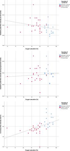
Patients with COVID-19 infection had a significantly lower hyperemic coronary flow velocity, resulting in a significantly lower CFVR (2.0 ± 0.3 vs. 2.4 ± 0.5, p < .001). Patients with severe COVID-19 had a lower CFVR compared to those with moderate COVID-19 (1.8 ± 0.2 vs. 2.2 ± 0.2, p < .001) driven by a trend toward higher basal flow velocity. CFVR correlated with troponin (p = .003, r: −.470), B-type natriuretic peptide (p < .001, r: −.580), C-reactive protein (p < .001, r: −.369), interleukin-6 (p < .001, r: −.597), and d-dimer (p < .001, r: −.561), with the three latter biomarkers having the highest areas-under-curve for predicting CMD.
Conclusions
Coronary microvascular dysfunction is common in patients with COVID-19 and is related to the severity of the infection. CMD may also explain the “cryptic” myocardial injury seen in patients with severe COVID-19 infection.
期刊介绍:
The journal features original contributions that are the result of investigations contributing significant new information relating to the vascular and lymphatic microcirculation addressed at the intact animal, organ, cellular, or molecular level. Papers describe applications of the methods of physiology, biophysics, bioengineering, genetics, cell biology, biochemistry, and molecular biology to problems in microcirculation.
Microcirculation also publishes state-of-the-art reviews that address frontier areas or new advances in technology in the fields of microcirculatory disease and function. Specific areas of interest include: Angiogenesis, growth and remodeling; Transport and exchange of gasses and solutes; Rheology and biorheology; Endothelial cell biology and metabolism; Interactions between endothelium, smooth muscle, parenchymal cells, leukocytes and platelets; Regulation of vasomotor tone; and Microvascular structures, imaging and morphometry. Papers also describe innovations in experimental techniques and instrumentation for studying all aspects of microcirculatory structure and function.


 分享
分享
 求助内容:
求助内容: 应助结果提醒方式:
 扫码关注我们
