Evropi Toulkeridou, Carlos Enrique Gutierrez, Daniel Baum, Kenji Doya, Evan P. Economo
{"title":"利用深度学习从微CT图像中自动分割昆虫解剖结构","authors":"Evropi Toulkeridou, Carlos Enrique Gutierrez, Daniel Baum, Kenji Doya, Evan P. Economo","doi":"10.1002/ntls.20230010","DOIUrl":null,"url":null,"abstract":"Abstract Three‐dimensional (3D) imaging, such as microcomputed tomography (micro‐CT), is increasingly being used by organismal biologists for precise and comprehensive anatomical characterization. However, the segmentation of anatomical structures remains a bottleneck in research, often requiring tedious manual work. Here, we propose a pipeline for the fully automated segmentation of anatomical structures in micro‐CT images utilizing state‐of‐the‐art deep learning methods, selecting the ant brain as a test case. We implemented the U‐Net architecture for two‐dimensional (2D) image segmentation for our convolutional neural network (CNN), combined with pixel‐island detection. For training and validation of the network, we assembled a data set of semimanually segmented brain images of 76 ant species. The trained network predicted the brain area in ant images fast and accurately; its performance tested on validation sets showed good agreement between the prediction and the target, scoring 80% Intersection over Union (IoU) and 90% Dice Coefficient (F1) accuracy. While manual segmentation usually takes many hours for each brain, the trained network takes only a few minutes. Furthermore, our network is generalizable for segmenting the whole neural system in full‐body scans, and works in tests on distantly related and morphologically divergent insects (e.g., fruit flies). The latter suggests that methods like the one presented here generally apply across diverse taxa. Our method makes the construction of segmented maps and the morphological quantification of different species more efficient and scalable to large data sets, a step toward a big data approach to organismal anatomy. Key points Development of a deep learning‐based pipeline for the fully automated segmentation of micro‐CT images of insects, using ant brains as a starting point. Creation of an open access data set of micro‐CT images of ant heads for training and testing. Generalizable computer vision methodology, extendable across diverse taxa and anatomical features.","PeriodicalId":74244,"journal":{"name":"Natural sciences (Weinheim, Germany)","volume":"124 1","pages":"0"},"PeriodicalIF":3.1000,"publicationDate":"2023-09-19","publicationTypes":"Journal Article","fieldsOfStudy":null,"isOpenAccess":false,"openAccessPdf":"","citationCount":"0","resultStr":"{\"title\":\"Automated segmentation of insect anatomy from micro‐CT images using deep learning\",\"authors\":\"Evropi Toulkeridou, Carlos Enrique Gutierrez, Daniel Baum, Kenji Doya, Evan P. Economo\",\"doi\":\"10.1002/ntls.20230010\",\"DOIUrl\":null,\"url\":null,\"abstract\":\"Abstract Three‐dimensional (3D) imaging, such as microcomputed tomography (micro‐CT), is increasingly being used by organismal biologists for precise and comprehensive anatomical characterization. However, the segmentation of anatomical structures remains a bottleneck in research, often requiring tedious manual work. Here, we propose a pipeline for the fully automated segmentation of anatomical structures in micro‐CT images utilizing state‐of‐the‐art deep learning methods, selecting the ant brain as a test case. We implemented the U‐Net architecture for two‐dimensional (2D) image segmentation for our convolutional neural network (CNN), combined with pixel‐island detection. For training and validation of the network, we assembled a data set of semimanually segmented brain images of 76 ant species. The trained network predicted the brain area in ant images fast and accurately; its performance tested on validation sets showed good agreement between the prediction and the target, scoring 80% Intersection over Union (IoU) and 90% Dice Coefficient (F1) accuracy. While manual segmentation usually takes many hours for each brain, the trained network takes only a few minutes. Furthermore, our network is generalizable for segmenting the whole neural system in full‐body scans, and works in tests on distantly related and morphologically divergent insects (e.g., fruit flies). The latter suggests that methods like the one presented here generally apply across diverse taxa. Our method makes the construction of segmented maps and the morphological quantification of different species more efficient and scalable to large data sets, a step toward a big data approach to organismal anatomy. Key points Development of a deep learning‐based pipeline for the fully automated segmentation of micro‐CT images of insects, using ant brains as a starting point. Creation of an open access data set of micro‐CT images of ant heads for training and testing. Generalizable computer vision methodology, extendable across diverse taxa and anatomical features.\",\"PeriodicalId\":74244,\"journal\":{\"name\":\"Natural sciences (Weinheim, Germany)\",\"volume\":\"124 1\",\"pages\":\"0\"},\"PeriodicalIF\":3.1000,\"publicationDate\":\"2023-09-19\",\"publicationTypes\":\"Journal Article\",\"fieldsOfStudy\":null,\"isOpenAccess\":false,\"openAccessPdf\":\"\",\"citationCount\":\"0\",\"resultStr\":null,\"platform\":\"Semanticscholar\",\"paperid\":null,\"PeriodicalName\":\"Natural sciences (Weinheim, Germany)\",\"FirstCategoryId\":\"1085\",\"ListUrlMain\":\"https://doi.org/10.1002/ntls.20230010\",\"RegionNum\":0,\"RegionCategory\":null,\"ArticlePicture\":[],\"TitleCN\":null,\"AbstractTextCN\":null,\"PMCID\":null,\"EPubDate\":\"\",\"PubModel\":\"\",\"JCR\":\"Q2\",\"JCRName\":\"MULTIDISCIPLINARY SCIENCES\",\"Score\":null,\"Total\":0}","platform":"Semanticscholar","paperid":null,"PeriodicalName":"Natural sciences (Weinheim, Germany)","FirstCategoryId":"1085","ListUrlMain":"https://doi.org/10.1002/ntls.20230010","RegionNum":0,"RegionCategory":null,"ArticlePicture":[],"TitleCN":null,"AbstractTextCN":null,"PMCID":null,"EPubDate":"","PubModel":"","JCR":"Q2","JCRName":"MULTIDISCIPLINARY SCIENCES","Score":null,"Total":0}
Automated segmentation of insect anatomy from micro‐CT images using deep learning
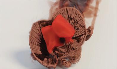
Abstract Three‐dimensional (3D) imaging, such as microcomputed tomography (micro‐CT), is increasingly being used by organismal biologists for precise and comprehensive anatomical characterization. However, the segmentation of anatomical structures remains a bottleneck in research, often requiring tedious manual work. Here, we propose a pipeline for the fully automated segmentation of anatomical structures in micro‐CT images utilizing state‐of‐the‐art deep learning methods, selecting the ant brain as a test case. We implemented the U‐Net architecture for two‐dimensional (2D) image segmentation for our convolutional neural network (CNN), combined with pixel‐island detection. For training and validation of the network, we assembled a data set of semimanually segmented brain images of 76 ant species. The trained network predicted the brain area in ant images fast and accurately; its performance tested on validation sets showed good agreement between the prediction and the target, scoring 80% Intersection over Union (IoU) and 90% Dice Coefficient (F1) accuracy. While manual segmentation usually takes many hours for each brain, the trained network takes only a few minutes. Furthermore, our network is generalizable for segmenting the whole neural system in full‐body scans, and works in tests on distantly related and morphologically divergent insects (e.g., fruit flies). The latter suggests that methods like the one presented here generally apply across diverse taxa. Our method makes the construction of segmented maps and the morphological quantification of different species more efficient and scalable to large data sets, a step toward a big data approach to organismal anatomy. Key points Development of a deep learning‐based pipeline for the fully automated segmentation of micro‐CT images of insects, using ant brains as a starting point. Creation of an open access data set of micro‐CT images of ant heads for training and testing. Generalizable computer vision methodology, extendable across diverse taxa and anatomical features.


 分享
分享

 求助内容:
求助内容: 应助结果提醒方式:
 扫码关注我们
