Samantha L. Gies, Maxx H. Tessmer, Dara W. Frank, Jimmy B. Feix
{"title":"对细菌磷脂酶 ExoU 的结构和膜相互作用的定点自旋标签 EPR 研究","authors":"Samantha L. Gies, Maxx H. Tessmer, Dara W. Frank, Jimmy B. Feix","doi":"10.1007/s00723-023-01620-0","DOIUrl":null,"url":null,"abstract":"<div><p>Site-directed spin labeling (SDSL) has been invaluable in the analysis of protein structure and dynamics and has been particularly useful in the study of membrane proteins. ExoU, an important virulence factor in <i>Pseudomonas aeruginosa</i> infections, is a bacterial phospholipase A2 that functions at the membrane—aqueous interface. Using the SDSL methodology developed in the Hubbell lab, we find that the region surrounding the catalytic site of ExoU is buried within the tertiary structure of the protein in the soluble, apoenzyme state, but shows a significant increase in dynamics upon membrane binding and activation by ubiquitin. Continuous wave (CW) power saturation EPR studies show that the conserved serine hydrolase motif of ExoU localizes to the membrane surface in the active, holoenzyme state. SDSL studies on the C-terminal four-helix bundle (4HB) domain of ExoU similarly show a co-operative effect of ubiquitin binding and membrane association. CW power saturation studies of the 4HB domain indicate that two interhelical loops intercalate into the lipid bilayer upon formation of the holoenzyme state, anchoring ExoU at the membrane surface. Together these studies establish the orientation and localization of ExoU at the membrane surface and illustrate the power of SDSL as applied to peripheral membrane proteins.</p></div>","PeriodicalId":469,"journal":{"name":"Applied Magnetic Resonance","volume":"55 1-3","pages":"279 - 295"},"PeriodicalIF":1.3000,"publicationDate":"2023-10-04","publicationTypes":"Journal Article","fieldsOfStudy":null,"isOpenAccess":false,"openAccessPdf":"","citationCount":"0","resultStr":"{\"title\":\"Site-Directed Spin Label EPR Studies of the Structure and Membrane Interactions of the Bacterial Phospholipase ExoU\",\"authors\":\"Samantha L. Gies, Maxx H. Tessmer, Dara W. Frank, Jimmy B. Feix\",\"doi\":\"10.1007/s00723-023-01620-0\",\"DOIUrl\":null,\"url\":null,\"abstract\":\"<div><p>Site-directed spin labeling (SDSL) has been invaluable in the analysis of protein structure and dynamics and has been particularly useful in the study of membrane proteins. ExoU, an important virulence factor in <i>Pseudomonas aeruginosa</i> infections, is a bacterial phospholipase A2 that functions at the membrane—aqueous interface. Using the SDSL methodology developed in the Hubbell lab, we find that the region surrounding the catalytic site of ExoU is buried within the tertiary structure of the protein in the soluble, apoenzyme state, but shows a significant increase in dynamics upon membrane binding and activation by ubiquitin. Continuous wave (CW) power saturation EPR studies show that the conserved serine hydrolase motif of ExoU localizes to the membrane surface in the active, holoenzyme state. SDSL studies on the C-terminal four-helix bundle (4HB) domain of ExoU similarly show a co-operative effect of ubiquitin binding and membrane association. CW power saturation studies of the 4HB domain indicate that two interhelical loops intercalate into the lipid bilayer upon formation of the holoenzyme state, anchoring ExoU at the membrane surface. Together these studies establish the orientation and localization of ExoU at the membrane surface and illustrate the power of SDSL as applied to peripheral membrane proteins.</p></div>\",\"PeriodicalId\":469,\"journal\":{\"name\":\"Applied Magnetic Resonance\",\"volume\":\"55 1-3\",\"pages\":\"279 - 295\"},\"PeriodicalIF\":1.3000,\"publicationDate\":\"2023-10-04\",\"publicationTypes\":\"Journal Article\",\"fieldsOfStudy\":null,\"isOpenAccess\":false,\"openAccessPdf\":\"\",\"citationCount\":\"0\",\"resultStr\":null,\"platform\":\"Semanticscholar\",\"paperid\":null,\"PeriodicalName\":\"Applied Magnetic Resonance\",\"FirstCategoryId\":\"101\",\"ListUrlMain\":\"https://link.springer.com/article/10.1007/s00723-023-01620-0\",\"RegionNum\":4,\"RegionCategory\":\"物理与天体物理\",\"ArticlePicture\":[],\"TitleCN\":null,\"AbstractTextCN\":null,\"PMCID\":null,\"EPubDate\":\"\",\"PubModel\":\"\",\"JCR\":\"Q4\",\"JCRName\":\"PHYSICS, ATOMIC, MOLECULAR & CHEMICAL\",\"Score\":null,\"Total\":0}","platform":"Semanticscholar","paperid":null,"PeriodicalName":"Applied Magnetic Resonance","FirstCategoryId":"101","ListUrlMain":"https://link.springer.com/article/10.1007/s00723-023-01620-0","RegionNum":4,"RegionCategory":"物理与天体物理","ArticlePicture":[],"TitleCN":null,"AbstractTextCN":null,"PMCID":null,"EPubDate":"","PubModel":"","JCR":"Q4","JCRName":"PHYSICS, ATOMIC, MOLECULAR & CHEMICAL","Score":null,"Total":0}
Site-Directed Spin Label EPR Studies of the Structure and Membrane Interactions of the Bacterial Phospholipase ExoU
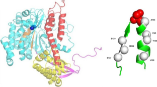
Site-directed spin labeling (SDSL) has been invaluable in the analysis of protein structure and dynamics and has been particularly useful in the study of membrane proteins. ExoU, an important virulence factor in Pseudomonas aeruginosa infections, is a bacterial phospholipase A2 that functions at the membrane—aqueous interface. Using the SDSL methodology developed in the Hubbell lab, we find that the region surrounding the catalytic site of ExoU is buried within the tertiary structure of the protein in the soluble, apoenzyme state, but shows a significant increase in dynamics upon membrane binding and activation by ubiquitin. Continuous wave (CW) power saturation EPR studies show that the conserved serine hydrolase motif of ExoU localizes to the membrane surface in the active, holoenzyme state. SDSL studies on the C-terminal four-helix bundle (4HB) domain of ExoU similarly show a co-operative effect of ubiquitin binding and membrane association. CW power saturation studies of the 4HB domain indicate that two interhelical loops intercalate into the lipid bilayer upon formation of the holoenzyme state, anchoring ExoU at the membrane surface. Together these studies establish the orientation and localization of ExoU at the membrane surface and illustrate the power of SDSL as applied to peripheral membrane proteins.
期刊介绍:
Applied Magnetic Resonance provides an international forum for the application of magnetic resonance in physics, chemistry, biology, medicine, geochemistry, ecology, engineering, and related fields.
The contents include articles with a strong emphasis on new applications, and on new experimental methods. Additional features include book reviews and Letters to the Editor.





 京公网安备 11010802042870号
京ICP备2023020795号-1
京公网安备 11010802042870号
京ICP备2023020795号-1

 分享
分享
 求助内容:
求助内容: 应助结果提醒方式:
 扫码关注我们
