{"title":"伴有钙化的颌下腺癌多形性腺瘤。","authors":"Tomoko Shiraishi, Toru Chikui, Shoko Yoshida, Kazuhiko Okamura, Kenichiro Hashimoto, Tomoko Fukui, Toyohiro Kagawa","doi":"10.1007/s11282-023-00724-4","DOIUrl":null,"url":null,"abstract":"<p><p>We report an unusual case of carcinoma ex pleomorphic adenoma (CXPA) in the submandibular gland. The mass had a unique calcification. Panoramic tomography revealed sponge-like calcification. The central portion displayed heterogeneous high signal intensity on T1-weighted image (T1WI) and T2-weighted image (T2WI), and heterogeneously moderate signal intensity on a short-TI inversion recovery (STIR) image. The ADC was low (0.78 × 10<sup>-3</sup>mm<sup>2</sup>/sec). After surgical excision, a pathological examination revealed that the mass contained CXPA as a minor component. Tumor cells with large hyperchromatic nuclei and eosinophilic or clear cytoplasm proliferated in irregular small tubule formations or cribriform or Roman-bridge structures in hyalinized or focally ossified stroma. The entire mass was calcified, particularly in the central region. Taken together, the reduced T1 relaxation times were related to the surface effects of diamagnetic particles, which were observed at calcium particle concentrations of up to 30%. We report a CXPA with unusual sponge-like calcification, which appeared unusually hyperintense on T1WI due to a surface effect.</p>","PeriodicalId":56103,"journal":{"name":"Oral Radiology","volume":" ","pages":"314-318"},"PeriodicalIF":1.6000,"publicationDate":"2024-04-01","publicationTypes":"Journal Article","fieldsOfStudy":null,"isOpenAccess":false,"openAccessPdf":"","citationCount":"0","resultStr":"{\"title\":\"Submandibular gland carcinoma ex pleomorphic adenoma with calcification.\",\"authors\":\"Tomoko Shiraishi, Toru Chikui, Shoko Yoshida, Kazuhiko Okamura, Kenichiro Hashimoto, Tomoko Fukui, Toyohiro Kagawa\",\"doi\":\"10.1007/s11282-023-00724-4\",\"DOIUrl\":null,\"url\":null,\"abstract\":\"<p><p>We report an unusual case of carcinoma ex pleomorphic adenoma (CXPA) in the submandibular gland. The mass had a unique calcification. Panoramic tomography revealed sponge-like calcification. The central portion displayed heterogeneous high signal intensity on T1-weighted image (T1WI) and T2-weighted image (T2WI), and heterogeneously moderate signal intensity on a short-TI inversion recovery (STIR) image. The ADC was low (0.78 × 10<sup>-3</sup>mm<sup>2</sup>/sec). After surgical excision, a pathological examination revealed that the mass contained CXPA as a minor component. Tumor cells with large hyperchromatic nuclei and eosinophilic or clear cytoplasm proliferated in irregular small tubule formations or cribriform or Roman-bridge structures in hyalinized or focally ossified stroma. The entire mass was calcified, particularly in the central region. Taken together, the reduced T1 relaxation times were related to the surface effects of diamagnetic particles, which were observed at calcium particle concentrations of up to 30%. We report a CXPA with unusual sponge-like calcification, which appeared unusually hyperintense on T1WI due to a surface effect.</p>\",\"PeriodicalId\":56103,\"journal\":{\"name\":\"Oral Radiology\",\"volume\":\" \",\"pages\":\"314-318\"},\"PeriodicalIF\":1.6000,\"publicationDate\":\"2024-04-01\",\"publicationTypes\":\"Journal Article\",\"fieldsOfStudy\":null,\"isOpenAccess\":false,\"openAccessPdf\":\"\",\"citationCount\":\"0\",\"resultStr\":null,\"platform\":\"Semanticscholar\",\"paperid\":null,\"PeriodicalName\":\"Oral Radiology\",\"FirstCategoryId\":\"3\",\"ListUrlMain\":\"https://doi.org/10.1007/s11282-023-00724-4\",\"RegionNum\":3,\"RegionCategory\":\"医学\",\"ArticlePicture\":[],\"TitleCN\":null,\"AbstractTextCN\":null,\"PMCID\":null,\"EPubDate\":\"2023/11/30 0:00:00\",\"PubModel\":\"Epub\",\"JCR\":\"Q3\",\"JCRName\":\"DENTISTRY, ORAL SURGERY & MEDICINE\",\"Score\":null,\"Total\":0}","platform":"Semanticscholar","paperid":null,"PeriodicalName":"Oral Radiology","FirstCategoryId":"3","ListUrlMain":"https://doi.org/10.1007/s11282-023-00724-4","RegionNum":3,"RegionCategory":"医学","ArticlePicture":[],"TitleCN":null,"AbstractTextCN":null,"PMCID":null,"EPubDate":"2023/11/30 0:00:00","PubModel":"Epub","JCR":"Q3","JCRName":"DENTISTRY, ORAL SURGERY & MEDICINE","Score":null,"Total":0}
Submandibular gland carcinoma ex pleomorphic adenoma with calcification.
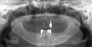
We report an unusual case of carcinoma ex pleomorphic adenoma (CXPA) in the submandibular gland. The mass had a unique calcification. Panoramic tomography revealed sponge-like calcification. The central portion displayed heterogeneous high signal intensity on T1-weighted image (T1WI) and T2-weighted image (T2WI), and heterogeneously moderate signal intensity on a short-TI inversion recovery (STIR) image. The ADC was low (0.78 × 10⁻³mm2/sec). After surgical excision, a pathological examination revealed that the mass contained CXPA as a minor component. Tumor cells with large hyperchromatic nuclei and eosinophilic or clear cytoplasm proliferated in irregular small tubule formations or cribriform or Roman-bridge structures in hyalinized or focally ossified stroma. The entire mass was calcified, particularly in the central region. Taken together, the reduced T1 relaxation times were related to the surface effects of diamagnetic particles, which were observed at calcium particle concentrations of up to 30%. We report a CXPA with unusual sponge-like calcification, which appeared unusually hyperintense on T1WI due to a surface effect.
期刊介绍:
As the official English-language journal of the Japanese Society for Oral and Maxillofacial Radiology and the Asian Academy of Oral and Maxillofacial Radiology, Oral Radiology is intended to be a forum for international collaboration in head and neck diagnostic imaging and all related fields. Oral Radiology features cutting-edge research papers, review articles, case reports, and technical notes from both the clinical and experimental fields. As membership in the Society is not a prerequisite, contributions are welcome from researchers and clinicians worldwide.


 分享
分享
 求助内容:
求助内容: 应助结果提醒方式:
 扫码关注我们
