Talal Bonny, Abdelaziz Al-Ali, Mohammed Al-Ali, Rashid Alsaadi, Wafaa Al Nassan, Khaled Obaideen, Maryam AlMallahi
{"title":"基于深度学习的卷积神经网络算法的牙咬翼x线片分割。","authors":"Talal Bonny, Abdelaziz Al-Ali, Mohammed Al-Ali, Rashid Alsaadi, Wafaa Al Nassan, Khaled Obaideen, Maryam AlMallahi","doi":"10.1007/s11282-023-00717-3","DOIUrl":null,"url":null,"abstract":"<p><strong>Objectives: </strong>Dental radiographs, particularly bitewing radiographs, are widely used in dental diagnosis and treatment Dental image segmentation is difficult for various reasons, such as intricate structures, low contrast, noise, roughness, and unclear borders, resulting in poor image quality. Recent developments in deep learning models have improved performance in analyzing dental images. In this research, our primary objective is to determine the most effective segmentation technique for bitewing radiographs based on different metrics: accuracy, training time, and the number of training parameters as a reflection of architectural cost.</p><p><strong>Methods: </strong>In this research, we employ several deep learning models, namely Resnet-18, Resnet-50, Xception, Inception Resnet v2, and Mobilenetv2, to segment bitewing radiographs. The process begins by importing the radiographs into MATLAB®(MathWorks Inc), where the images are first improved, then segmented using the graph cut method based on regions to produce a binary mask that distinguishes the background from the original X-ray.</p><p><strong>Results: </strong>The deep learning models were trained on 298 and 99 radiograph training and validation sets and were evaluated using 99 images from the testing set. We also compare the segmentation model using several criteria, including accuracy, speed, and size, to determine which network is superior. Furthermore, we compare our findings with prior research to provide a comprehensive understanding of the advancements made in dental image segmentation. The accurate segmentation achieved was 93.67% and 94.42% by the Resnet-18 and Resnet-50 models, respectively.</p><p><strong>Conclusion: </strong>This research advances dental image analysis and facilitates more accurate diagnoses and treatment planning by determining the best segmentation technique. The outcomes of this study can guide researchers and practitioners in selecting appropriate segmentation methods for practical dental image analysis.</p>","PeriodicalId":56103,"journal":{"name":"Oral Radiology","volume":" ","pages":"165-177"},"PeriodicalIF":1.7000,"publicationDate":"2024-04-01","publicationTypes":"Journal Article","fieldsOfStudy":null,"isOpenAccess":false,"openAccessPdf":"","citationCount":"0","resultStr":"{\"title\":\"Dental bitewing radiographs segmentation using deep learning-based convolutional neural network algorithms.\",\"authors\":\"Talal Bonny, Abdelaziz Al-Ali, Mohammed Al-Ali, Rashid Alsaadi, Wafaa Al Nassan, Khaled Obaideen, Maryam AlMallahi\",\"doi\":\"10.1007/s11282-023-00717-3\",\"DOIUrl\":null,\"url\":null,\"abstract\":\"<p><strong>Objectives: </strong>Dental radiographs, particularly bitewing radiographs, are widely used in dental diagnosis and treatment Dental image segmentation is difficult for various reasons, such as intricate structures, low contrast, noise, roughness, and unclear borders, resulting in poor image quality. Recent developments in deep learning models have improved performance in analyzing dental images. In this research, our primary objective is to determine the most effective segmentation technique for bitewing radiographs based on different metrics: accuracy, training time, and the number of training parameters as a reflection of architectural cost.</p><p><strong>Methods: </strong>In this research, we employ several deep learning models, namely Resnet-18, Resnet-50, Xception, Inception Resnet v2, and Mobilenetv2, to segment bitewing radiographs. The process begins by importing the radiographs into MATLAB®(MathWorks Inc), where the images are first improved, then segmented using the graph cut method based on regions to produce a binary mask that distinguishes the background from the original X-ray.</p><p><strong>Results: </strong>The deep learning models were trained on 298 and 99 radiograph training and validation sets and were evaluated using 99 images from the testing set. We also compare the segmentation model using several criteria, including accuracy, speed, and size, to determine which network is superior. Furthermore, we compare our findings with prior research to provide a comprehensive understanding of the advancements made in dental image segmentation. The accurate segmentation achieved was 93.67% and 94.42% by the Resnet-18 and Resnet-50 models, respectively.</p><p><strong>Conclusion: </strong>This research advances dental image analysis and facilitates more accurate diagnoses and treatment planning by determining the best segmentation technique. The outcomes of this study can guide researchers and practitioners in selecting appropriate segmentation methods for practical dental image analysis.</p>\",\"PeriodicalId\":56103,\"journal\":{\"name\":\"Oral Radiology\",\"volume\":\" \",\"pages\":\"165-177\"},\"PeriodicalIF\":1.7000,\"publicationDate\":\"2024-04-01\",\"publicationTypes\":\"Journal Article\",\"fieldsOfStudy\":null,\"isOpenAccess\":false,\"openAccessPdf\":\"\",\"citationCount\":\"0\",\"resultStr\":null,\"platform\":\"Semanticscholar\",\"paperid\":null,\"PeriodicalName\":\"Oral Radiology\",\"FirstCategoryId\":\"3\",\"ListUrlMain\":\"https://doi.org/10.1007/s11282-023-00717-3\",\"RegionNum\":3,\"RegionCategory\":\"医学\",\"ArticlePicture\":[],\"TitleCN\":null,\"AbstractTextCN\":null,\"PMCID\":null,\"EPubDate\":\"2023/12/4 0:00:00\",\"PubModel\":\"Epub\",\"JCR\":\"Q3\",\"JCRName\":\"DENTISTRY, ORAL SURGERY & MEDICINE\",\"Score\":null,\"Total\":0}","platform":"Semanticscholar","paperid":null,"PeriodicalName":"Oral Radiology","FirstCategoryId":"3","ListUrlMain":"https://doi.org/10.1007/s11282-023-00717-3","RegionNum":3,"RegionCategory":"医学","ArticlePicture":[],"TitleCN":null,"AbstractTextCN":null,"PMCID":null,"EPubDate":"2023/12/4 0:00:00","PubModel":"Epub","JCR":"Q3","JCRName":"DENTISTRY, ORAL SURGERY & MEDICINE","Score":null,"Total":0}
Dental bitewing radiographs segmentation using deep learning-based convolutional neural network algorithms.
Objectives: Dental radiographs, particularly bitewing radiographs, are widely used in dental diagnosis and treatment Dental image segmentation is difficult for various reasons, such as intricate structures, low contrast, noise, roughness, and unclear borders, resulting in poor image quality. Recent developments in deep learning models have improved performance in analyzing dental images. In this research, our primary objective is to determine the most effective segmentation technique for bitewing radiographs based on different metrics: accuracy, training time, and the number of training parameters as a reflection of architectural cost.
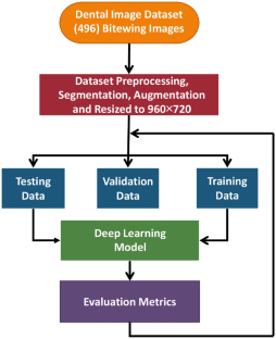
Methods: In this research, we employ several deep learning models, namely Resnet-18, Resnet-50, Xception, Inception Resnet v2, and Mobilenetv2, to segment bitewing radiographs. The process begins by importing the radiographs into MATLAB®(MathWorks Inc), where the images are first improved, then segmented using the graph cut method based on regions to produce a binary mask that distinguishes the background from the original X-ray.
Results: The deep learning models were trained on 298 and 99 radiograph training and validation sets and were evaluated using 99 images from the testing set. We also compare the segmentation model using several criteria, including accuracy, speed, and size, to determine which network is superior. Furthermore, we compare our findings with prior research to provide a comprehensive understanding of the advancements made in dental image segmentation. The accurate segmentation achieved was 93.67% and 94.42% by the Resnet-18 and Resnet-50 models, respectively.
Conclusion: This research advances dental image analysis and facilitates more accurate diagnoses and treatment planning by determining the best segmentation technique. The outcomes of this study can guide researchers and practitioners in selecting appropriate segmentation methods for practical dental image analysis.
期刊介绍:
As the official English-language journal of the Japanese Society for Oral and Maxillofacial Radiology and the Asian Academy of Oral and Maxillofacial Radiology, Oral Radiology is intended to be a forum for international collaboration in head and neck diagnostic imaging and all related fields. Oral Radiology features cutting-edge research papers, review articles, case reports, and technical notes from both the clinical and experimental fields. As membership in the Society is not a prerequisite, contributions are welcome from researchers and clinicians worldwide.


 分享
分享
 求助内容:
求助内容: 应助结果提醒方式:
 扫码关注我们
