Hong Ki Min, Se Hee Kim, Hae-Rim Kim, Sang-Heon Lee
{"title":"强直性脊柱炎患者齿状突孤立综合征1例。","authors":"Hong Ki Min, Se Hee Kim, Hae-Rim Kim, Sang-Heon Lee","doi":"10.46497/ArchRheumatol.2023.9880","DOIUrl":null,"url":null,"abstract":"A 22-year-old male patient was admitted to the rheumatology clinic due to neck and back pain with morning stiffness for 12 months. He suffered from right knee and heel pain six months ago. He showed increased levels of high-sensitivity C-reactive protein (2.20 mg/dL, reference range: 0.01-0.3 mg/dL) and erythrocyte sedimentation rate (67 mm/h, reference range: 0-15 mm/h). To evaluate neck and back pain, the physician performed computed tomography (CT) of the whole spine. The CT finding was absence for herniated nucleus pulposus nor neural foramen stenosis. All vertebral corners of whole spine were clear (Figure 1a), except for bony spur at odontoid process of C2 (Figure 1b, white arrow).","PeriodicalId":93884,"journal":{"name":"Archives of rheumatology","volume":"38 3","pages":"480-481"},"PeriodicalIF":1.1000,"publicationDate":"2022-11-04","publicationTypes":"Journal Article","fieldsOfStudy":null,"isOpenAccess":false,"openAccessPdf":"https://www.ncbi.nlm.nih.gov/pmc/articles/PMC10689010/pdf/","citationCount":"0","resultStr":"{\"title\":\"Solitary syndesmophyte in odontoid process of a patient with ankylosing spondylitis.\",\"authors\":\"Hong Ki Min, Se Hee Kim, Hae-Rim Kim, Sang-Heon Lee\",\"doi\":\"10.46497/ArchRheumatol.2023.9880\",\"DOIUrl\":null,\"url\":null,\"abstract\":\"A 22-year-old male patient was admitted to the rheumatology clinic due to neck and back pain with morning stiffness for 12 months. He suffered from right knee and heel pain six months ago. He showed increased levels of high-sensitivity C-reactive protein (2.20 mg/dL, reference range: 0.01-0.3 mg/dL) and erythrocyte sedimentation rate (67 mm/h, reference range: 0-15 mm/h). To evaluate neck and back pain, the physician performed computed tomography (CT) of the whole spine. The CT finding was absence for herniated nucleus pulposus nor neural foramen stenosis. All vertebral corners of whole spine were clear (Figure 1a), except for bony spur at odontoid process of C2 (Figure 1b, white arrow).\",\"PeriodicalId\":93884,\"journal\":{\"name\":\"Archives of rheumatology\",\"volume\":\"38 3\",\"pages\":\"480-481\"},\"PeriodicalIF\":1.1000,\"publicationDate\":\"2022-11-04\",\"publicationTypes\":\"Journal Article\",\"fieldsOfStudy\":null,\"isOpenAccess\":false,\"openAccessPdf\":\"https://www.ncbi.nlm.nih.gov/pmc/articles/PMC10689010/pdf/\",\"citationCount\":\"0\",\"resultStr\":null,\"platform\":\"Semanticscholar\",\"paperid\":null,\"PeriodicalName\":\"Archives of rheumatology\",\"FirstCategoryId\":\"1085\",\"ListUrlMain\":\"https://doi.org/10.46497/ArchRheumatol.2023.9880\",\"RegionNum\":0,\"RegionCategory\":null,\"ArticlePicture\":[],\"TitleCN\":null,\"AbstractTextCN\":null,\"PMCID\":null,\"EPubDate\":\"2023/9/1 0:00:00\",\"PubModel\":\"eCollection\",\"JCR\":\"Q4\",\"JCRName\":\"RHEUMATOLOGY\",\"Score\":null,\"Total\":0}","platform":"Semanticscholar","paperid":null,"PeriodicalName":"Archives of rheumatology","FirstCategoryId":"1085","ListUrlMain":"https://doi.org/10.46497/ArchRheumatol.2023.9880","RegionNum":0,"RegionCategory":null,"ArticlePicture":[],"TitleCN":null,"AbstractTextCN":null,"PMCID":null,"EPubDate":"2023/9/1 0:00:00","PubModel":"eCollection","JCR":"Q4","JCRName":"RHEUMATOLOGY","Score":null,"Total":0}
Solitary syndesmophyte in odontoid process of a patient with ankylosing spondylitis.
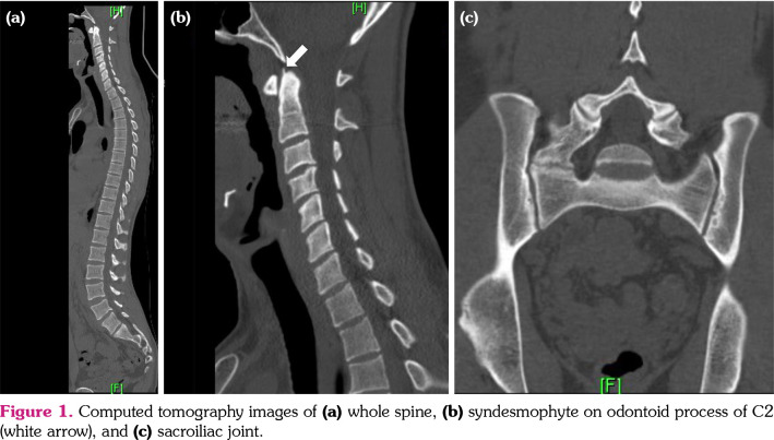
A 22-year-old male patient was admitted to the rheumatology clinic due to neck and back pain with morning stiffness for 12 months. He suffered from right knee and heel pain six months ago. He showed increased levels of high-sensitivity C-reactive protein (2.20 mg/dL, reference range: 0.01-0.3 mg/dL) and erythrocyte sedimentation rate (67 mm/h, reference range: 0-15 mm/h). To evaluate neck and back pain, the physician performed computed tomography (CT) of the whole spine. The CT finding was absence for herniated nucleus pulposus nor neural foramen stenosis. All vertebral corners of whole spine were clear (Figure 1a), except for bony spur at odontoid process of C2 (Figure 1b, white arrow).


 分享
分享

 求助内容:
求助内容: 应助结果提醒方式:
 扫码关注我们
