E. G. Novoselova, O. V. Glushkova, M. O. Khrenov, S. M. Lunin, M. G. Sharapov, R. G. Goncharov, E. K. Mubarakshina, T. V. Novoselova, S. B. Parfenyuk
{"title":"胸腺激素胸腺素-1α可降低内毒素诱导的生264.7细胞的促炎反应","authors":"E. G. Novoselova, O. V. Glushkova, M. O. Khrenov, S. M. Lunin, M. G. Sharapov, R. G. Goncharov, E. K. Mubarakshina, T. V. Novoselova, S. B. Parfenyuk","doi":"10.1134/s0026893323060110","DOIUrl":null,"url":null,"abstract":"<p><b>Abstract</b>—The aim of this work was to study the effects of thymosin-1 alpha (Tα1) on the anti-inflammatory response of RAW 264.7 macrophages cultured in the presence of lipopolysaccharide (LPS) from the walls of gram-negative bacteria. As well, we evaluated production of pro-inflammatory cytokines and the activity of the NF-κB and SAPK/JNK signaling pathways. In addition, the level of expression of a number of genes that regulate cell apoptosis, as well as the activity of receptors involved in the pro-inflammatory response, was determined. First, the addition of Tα1 normalized the level of cytokine production to varying degrees, with a particularly noticeable effect on IL-1β and IL-6. Second, the addition of Tα1 normalized the activity of the NF-κB and SAPK/JNK signaling cascades and the expression of the <i>Tlr4</i> gene. Third, Tα1 significantly reduced p53 and the activity of the <i>P53</i> gene, which is a marker of cell apoptosis. Fourth, it was shown that the increase in <i>Ar-1</i> gene expression under the influence of LPS was significantly reduced using Tα1. Thus, it was found that the presence of Tα1 in the RAW 264.7 cell culture medium significantly reduced the level of the pro-inflammatory response of cells.</p>","PeriodicalId":18734,"journal":{"name":"Molecular Biology","volume":"68 1","pages":""},"PeriodicalIF":1.2000,"publicationDate":"2023-12-07","publicationTypes":"Journal Article","fieldsOfStudy":null,"isOpenAccess":false,"openAccessPdf":"","citationCount":"0","resultStr":"{\"title\":\"The Thymic Hormone Thymosin-1α Reduces the Pro-Inflammatory Response of Raw 264.7 Cells Induced by Endotoxin\",\"authors\":\"E. G. Novoselova, O. V. Glushkova, M. O. Khrenov, S. M. Lunin, M. G. Sharapov, R. G. Goncharov, E. K. Mubarakshina, T. V. Novoselova, S. B. Parfenyuk\",\"doi\":\"10.1134/s0026893323060110\",\"DOIUrl\":null,\"url\":null,\"abstract\":\"<p><b>Abstract</b>—The aim of this work was to study the effects of thymosin-1 alpha (Tα1) on the anti-inflammatory response of RAW 264.7 macrophages cultured in the presence of lipopolysaccharide (LPS) from the walls of gram-negative bacteria. As well, we evaluated production of pro-inflammatory cytokines and the activity of the NF-κB and SAPK/JNK signaling pathways. In addition, the level of expression of a number of genes that regulate cell apoptosis, as well as the activity of receptors involved in the pro-inflammatory response, was determined. First, the addition of Tα1 normalized the level of cytokine production to varying degrees, with a particularly noticeable effect on IL-1β and IL-6. Second, the addition of Tα1 normalized the activity of the NF-κB and SAPK/JNK signaling cascades and the expression of the <i>Tlr4</i> gene. Third, Tα1 significantly reduced p53 and the activity of the <i>P53</i> gene, which is a marker of cell apoptosis. Fourth, it was shown that the increase in <i>Ar-1</i> gene expression under the influence of LPS was significantly reduced using Tα1. Thus, it was found that the presence of Tα1 in the RAW 264.7 cell culture medium significantly reduced the level of the pro-inflammatory response of cells.</p>\",\"PeriodicalId\":18734,\"journal\":{\"name\":\"Molecular Biology\",\"volume\":\"68 1\",\"pages\":\"\"},\"PeriodicalIF\":1.2000,\"publicationDate\":\"2023-12-07\",\"publicationTypes\":\"Journal Article\",\"fieldsOfStudy\":null,\"isOpenAccess\":false,\"openAccessPdf\":\"\",\"citationCount\":\"0\",\"resultStr\":null,\"platform\":\"Semanticscholar\",\"paperid\":null,\"PeriodicalName\":\"Molecular Biology\",\"FirstCategoryId\":\"99\",\"ListUrlMain\":\"https://doi.org/10.1134/s0026893323060110\",\"RegionNum\":4,\"RegionCategory\":\"生物学\",\"ArticlePicture\":[],\"TitleCN\":null,\"AbstractTextCN\":null,\"PMCID\":null,\"EPubDate\":\"\",\"PubModel\":\"\",\"JCR\":\"Q4\",\"JCRName\":\"BIOCHEMISTRY & MOLECULAR BIOLOGY\",\"Score\":null,\"Total\":0}","platform":"Semanticscholar","paperid":null,"PeriodicalName":"Molecular Biology","FirstCategoryId":"99","ListUrlMain":"https://doi.org/10.1134/s0026893323060110","RegionNum":4,"RegionCategory":"生物学","ArticlePicture":[],"TitleCN":null,"AbstractTextCN":null,"PMCID":null,"EPubDate":"","PubModel":"","JCR":"Q4","JCRName":"BIOCHEMISTRY & MOLECULAR BIOLOGY","Score":null,"Total":0}
The Thymic Hormone Thymosin-1α Reduces the Pro-Inflammatory Response of Raw 264.7 Cells Induced by Endotoxin
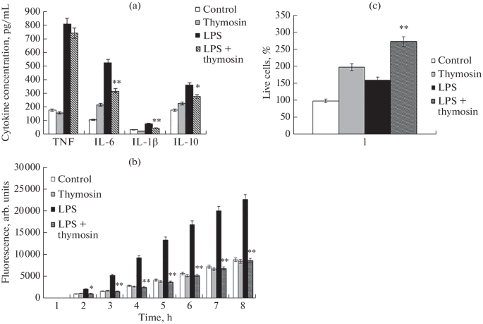
Abstract—The aim of this work was to study the effects of thymosin-1 alpha (Tα1) on the anti-inflammatory response of RAW 264.7 macrophages cultured in the presence of lipopolysaccharide (LPS) from the walls of gram-negative bacteria. As well, we evaluated production of pro-inflammatory cytokines and the activity of the NF-κB and SAPK/JNK signaling pathways. In addition, the level of expression of a number of genes that regulate cell apoptosis, as well as the activity of receptors involved in the pro-inflammatory response, was determined. First, the addition of Tα1 normalized the level of cytokine production to varying degrees, with a particularly noticeable effect on IL-1β and IL-6. Second, the addition of Tα1 normalized the activity of the NF-κB and SAPK/JNK signaling cascades and the expression of the Tlr4 gene. Third, Tα1 significantly reduced p53 and the activity of the P53 gene, which is a marker of cell apoptosis. Fourth, it was shown that the increase in Ar-1 gene expression under the influence of LPS was significantly reduced using Tα1. Thus, it was found that the presence of Tα1 in the RAW 264.7 cell culture medium significantly reduced the level of the pro-inflammatory response of cells.
期刊介绍:
Molecular Biology is an international peer reviewed journal that covers a wide scope of problems in molecular, cell and computational biology including genomics, proteomics, bioinformatics, molecular virology and immunology, molecular development biology, molecular evolution and related areals. Molecular Biology publishes reviews, experimental and theoretical works. Every year, the journal publishes special issues devoted to most rapidly developing branches of physical-chemical biology and to the most outstanding scientists.


 分享
分享
 求助内容:
求助内容: 应助结果提醒方式:
 扫码关注我们
