Preetha Krishnamurthy, Uma Maheswari, Kasim Mohamed
{"title":"假体康复对无牙颌阻塞性睡眠呼吸暂停患者气道空间的影响--一项初步观察研究。","authors":"Preetha Krishnamurthy, Uma Maheswari, Kasim Mohamed","doi":"10.30476/dentjods.2022.95716.1886","DOIUrl":null,"url":null,"abstract":"<p><strong>Statement of the problem: </strong>The common causes of obstructive sleep apnea (OSA) are identified as anatomic and/or functional abnormality in the oral cavity, oropharynx, velopharynx, and hypopharynx leading to compromised airway space and increased collapsibility.</p><p><strong>Purpose: </strong>This study was conducted to evaluate the effect of implant-supported mandibular complete denture in improving the airway space among completely edentulous patients with OSA and compare it with conventional complete denture.</p><p><strong>Materials and method: </strong>In this observational study, completely edentulous individuals were screened with snoring, tiredness, observed apnea, high blood pressure, body mass index, age, neck circumference, and gender (STOP-Bang) questionnaire to evaluate the incidence of OSA. Ten mild-moderate patients were included as study participants. Lateral cephalograms (L1) made at the edentulous state was considered baseline. They were rehabilitated with complete denture prosthesis. One week after denture insertion, two implants were placed in the edentulous mandibular arch. Delayed loading protocol was followed. Lateral cephalogram (L2) was made 6 months after complete denture insertion and 6 months after implant-supported prosthesis (L3). Cephalometric tracings were used to evaluate change in upper airway space (UAS), middle airway space (MAS), and lower airway space (LAS). Repeated measures ANOVA was used to evaluate statistical significance in the airway measurements made at the three intervals. Post hoc Tukey HSD and Bonferroni test were used to assess if the differences obtained were truly significant.</p><p><strong>Results: </strong>Statistical analysis revealed significant differences in UAS, MAS and LAS between L1, L2 and L3 (<i>p</i>< 0.05). Post hoc Tukey HSD indicated that UAS increased significantly at all three intervals followed by LAS and MAS respectively (α=.05). Post hoc Bon-ferroni test indicated that implant-supported mandibular complete dentures had a significant improvement in airway space when compared to conventional complete dentures (α=.05).</p><p><strong>Conclusion: </strong>Implant-supported mandibular complete denture could be effective in edentulous patients with mild-moderate OSA.</p>","PeriodicalId":73702,"journal":{"name":"Journal of dentistry (Shiraz, Iran)","volume":"24 4","pages":"382-388"},"PeriodicalIF":0.0000,"publicationDate":"2023-12-01","publicationTypes":"Journal Article","fieldsOfStudy":null,"isOpenAccess":false,"openAccessPdf":"https://www.ncbi.nlm.nih.gov/pmc/articles/PMC10749438/pdf/","citationCount":"0","resultStr":"{\"title\":\"Effect of Prosthetic Rehabilitation on Airway Space in Edentulous Patients with Obstructive Sleep Apnea- a Preliminary Observational Study.\",\"authors\":\"Preetha Krishnamurthy, Uma Maheswari, Kasim Mohamed\",\"doi\":\"10.30476/dentjods.2022.95716.1886\",\"DOIUrl\":null,\"url\":null,\"abstract\":\"<p><strong>Statement of the problem: </strong>The common causes of obstructive sleep apnea (OSA) are identified as anatomic and/or functional abnormality in the oral cavity, oropharynx, velopharynx, and hypopharynx leading to compromised airway space and increased collapsibility.</p><p><strong>Purpose: </strong>This study was conducted to evaluate the effect of implant-supported mandibular complete denture in improving the airway space among completely edentulous patients with OSA and compare it with conventional complete denture.</p><p><strong>Materials and method: </strong>In this observational study, completely edentulous individuals were screened with snoring, tiredness, observed apnea, high blood pressure, body mass index, age, neck circumference, and gender (STOP-Bang) questionnaire to evaluate the incidence of OSA. Ten mild-moderate patients were included as study participants. Lateral cephalograms (L1) made at the edentulous state was considered baseline. They were rehabilitated with complete denture prosthesis. One week after denture insertion, two implants were placed in the edentulous mandibular arch. Delayed loading protocol was followed. Lateral cephalogram (L2) was made 6 months after complete denture insertion and 6 months after implant-supported prosthesis (L3). Cephalometric tracings were used to evaluate change in upper airway space (UAS), middle airway space (MAS), and lower airway space (LAS). Repeated measures ANOVA was used to evaluate statistical significance in the airway measurements made at the three intervals. Post hoc Tukey HSD and Bonferroni test were used to assess if the differences obtained were truly significant.</p><p><strong>Results: </strong>Statistical analysis revealed significant differences in UAS, MAS and LAS between L1, L2 and L3 (<i>p</i>< 0.05). Post hoc Tukey HSD indicated that UAS increased significantly at all three intervals followed by LAS and MAS respectively (α=.05). Post hoc Bon-ferroni test indicated that implant-supported mandibular complete dentures had a significant improvement in airway space when compared to conventional complete dentures (α=.05).</p><p><strong>Conclusion: </strong>Implant-supported mandibular complete denture could be effective in edentulous patients with mild-moderate OSA.</p>\",\"PeriodicalId\":73702,\"journal\":{\"name\":\"Journal of dentistry (Shiraz, Iran)\",\"volume\":\"24 4\",\"pages\":\"382-388\"},\"PeriodicalIF\":0.0000,\"publicationDate\":\"2023-12-01\",\"publicationTypes\":\"Journal Article\",\"fieldsOfStudy\":null,\"isOpenAccess\":false,\"openAccessPdf\":\"https://www.ncbi.nlm.nih.gov/pmc/articles/PMC10749438/pdf/\",\"citationCount\":\"0\",\"resultStr\":null,\"platform\":\"Semanticscholar\",\"paperid\":null,\"PeriodicalName\":\"Journal of dentistry (Shiraz, Iran)\",\"FirstCategoryId\":\"1085\",\"ListUrlMain\":\"https://doi.org/10.30476/dentjods.2022.95716.1886\",\"RegionNum\":0,\"RegionCategory\":null,\"ArticlePicture\":[],\"TitleCN\":null,\"AbstractTextCN\":null,\"PMCID\":null,\"EPubDate\":\"\",\"PubModel\":\"\",\"JCR\":\"\",\"JCRName\":\"\",\"Score\":null,\"Total\":0}","platform":"Semanticscholar","paperid":null,"PeriodicalName":"Journal of dentistry (Shiraz, Iran)","FirstCategoryId":"1085","ListUrlMain":"https://doi.org/10.30476/dentjods.2022.95716.1886","RegionNum":0,"RegionCategory":null,"ArticlePicture":[],"TitleCN":null,"AbstractTextCN":null,"PMCID":null,"EPubDate":"","PubModel":"","JCR":"","JCRName":"","Score":null,"Total":0}
引用次数: 0
摘要
问题简介:阻塞性睡眠呼吸暂停(OSA)的常见原因被认为是口腔、口咽、咽后部和咽下部的解剖和/或功能异常导致气道空间受损和塌陷增加:在这项观察性研究中,对全口无牙患者进行打鼾、疲倦、观察到的呼吸暂停、高血压、体重指数、年龄、颈围和性别(STOP-Bang)问卷调查,以评估 OSA 的发生率。十名轻中度患者被纳入研究对象。无牙颌状态下的侧位头影(L1)被视为基线。他们接受了全口义齿修复。安装义齿一周后,在无牙颌的下颌弓上植入两颗种植体。采用延迟加载方案。在安装全口义齿 6 个月后和种植体支持修复体(L3)6 个月后分别进行了侧位头影(L2)检查。头颅测量描记图用于评估上气道间隙(UAS)、中气道间隙(MAS)和下气道间隙(LAS)的变化。采用重复测量方差分析来评估三个间隔期气道测量的统计学意义。采用事后 Tukey HSD 和 Bonferroni 检验来评估所获得的差异是否真正显著:统计分析显示,L1、L2 和 L3 之间的 UAS、MAS 和 LAS 存在明显差异(p< 0.05)。事后 Tukey HSD 表明,UAS 在所有三个时间间隔都有明显增加,其次分别是 LAS 和 MAS(α=.05)。事后 Bon-ferroni 检验表明,与传统全口义齿相比,种植体支持的下颌全口义齿在气道空间方面有明显改善(α=.05):结论:种植体支持下颌全口义齿对无牙颌的轻中度 OSA 患者有效。
Effect of Prosthetic Rehabilitation on Airway Space in Edentulous Patients with Obstructive Sleep Apnea- a Preliminary Observational Study.
Statement of the problem: The common causes of obstructive sleep apnea (OSA) are identified as anatomic and/or functional abnormality in the oral cavity, oropharynx, velopharynx, and hypopharynx leading to compromised airway space and increased collapsibility.
Purpose: This study was conducted to evaluate the effect of implant-supported mandibular complete denture in improving the airway space among completely edentulous patients with OSA and compare it with conventional complete denture.
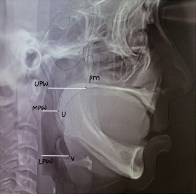
Materials and method: In this observational study, completely edentulous individuals were screened with snoring, tiredness, observed apnea, high blood pressure, body mass index, age, neck circumference, and gender (STOP-Bang) questionnaire to evaluate the incidence of OSA. Ten mild-moderate patients were included as study participants. Lateral cephalograms (L1) made at the edentulous state was considered baseline. They were rehabilitated with complete denture prosthesis. One week after denture insertion, two implants were placed in the edentulous mandibular arch. Delayed loading protocol was followed. Lateral cephalogram (L2) was made 6 months after complete denture insertion and 6 months after implant-supported prosthesis (L3). Cephalometric tracings were used to evaluate change in upper airway space (UAS), middle airway space (MAS), and lower airway space (LAS). Repeated measures ANOVA was used to evaluate statistical significance in the airway measurements made at the three intervals. Post hoc Tukey HSD and Bonferroni test were used to assess if the differences obtained were truly significant.
Results: Statistical analysis revealed significant differences in UAS, MAS and LAS between L1, L2 and L3 (p< 0.05). Post hoc Tukey HSD indicated that UAS increased significantly at all three intervals followed by LAS and MAS respectively (α=.05). Post hoc Bon-ferroni test indicated that implant-supported mandibular complete dentures had a significant improvement in airway space when compared to conventional complete dentures (α=.05).
Conclusion: Implant-supported mandibular complete denture could be effective in edentulous patients with mild-moderate OSA.


 分享
分享

 求助内容:
求助内容: 应助结果提醒方式:
 扫码关注我们
