{"title":"在小鸡胚胎白内障实验模型中研究蛹虫草苷的抗氧化作用。","authors":"Gülan Albaş Kurt, Tolga Ertekin, Emre Atay, Abdülkadir Bilir, Halit Buğra Koca, Esra Aslan, Alperen Sarıtaş","doi":"","DOIUrl":null,"url":null,"abstract":"<p><strong>Purpose: </strong>Cataract, which occurs as a result of lens opacification, is one of the most common causes of vision loss. In the literature, deterioration of the antioxidant system due to the increase in reactive oxygen species and oxidant levels is shown among the causes of cataract formation. The aim of this study was to investigate the antioxidant effect of chrysin on steroid-induced cataract development in an experimental chick embryo model using morphological, histological and biochemical parameters.</p><p><strong>Methods: </strong>Within the scope of the study, 150 specific pathogen free (SPF) fertilized eggs were used. Eggs were divided into 6 groups as control (group 1), corn oil (group 2), hydrocortisone hemisuccinate sodium (HC) (group 3), low dose chrysin (group 4), medium dose chrysin (group 5) and high dose chrysin (group 6). On the 15th day of incubation, Chrysin and HC were applicated to the air sac of the eggs with Hamilton and/or insulin injector. On day 17, the chick embryos were removed from the eggs and the bulbus oculi of the embryos were dissected. Lenses of 9 embryos were used for morpholigical cataract grading in each group, lens of 8 embryos for biochemical analysis and intact eyes of 7 embryos for histological evaluation (TUNEL method).</p><p><strong>Results: </strong>No opacity was observed in any of the lenses in Group 1 and 2. Cataract was observed in all lenses in Group 3. The mean opacity grades in group 3 were statistically significantly higher when compared to group 1 and 2 (p<0.05). The difference between group 6 and group 3 was statistically significant (p<0.05). GSH and TAS levels in the lenses were statistically significantly decreased compared to the control group due to HC application (p<0.05). It was determined that the decreased GSH and TAS levels in the lenses increased in relation to the Chrysin application doses. The increased levels of MDA, TOS, caspase 3 and caspase 9 in the HC group decreased significantly depending to the chrysin doses (p<0.05). In addition, while the rate of apoptotic cells determined by the TUNEL method was statistically significantly higher in the HC administered group than in the control group (p<0.05), it was statistically significantly decreased in the chrysin-administered groups, in relation to the dose of chrysin (p<0.05).</p><p><strong>Conclusions: </strong>We think that anti-cataract effect of crhysin may be due to the antioxidant and antiapoptotic properties of chrysin. However, more research is needed to clarify the anti-cataract effects of chrysin.</p>","PeriodicalId":18866,"journal":{"name":"Molecular Vision","volume":"29 ","pages":"245-255"},"PeriodicalIF":1.4000,"publicationDate":"2023-11-05","publicationTypes":"Journal Article","fieldsOfStudy":null,"isOpenAccess":false,"openAccessPdf":"https://www.ncbi.nlm.nih.gov/pmc/articles/PMC10784222/pdf/","citationCount":"0","resultStr":"{\"title\":\"Investigation of the antioxidant effect of Chrysin in an experimental cataract model created in chick embryos.\",\"authors\":\"Gülan Albaş Kurt, Tolga Ertekin, Emre Atay, Abdülkadir Bilir, Halit Buğra Koca, Esra Aslan, Alperen Sarıtaş\",\"doi\":\"\",\"DOIUrl\":null,\"url\":null,\"abstract\":\"<p><strong>Purpose: </strong>Cataract, which occurs as a result of lens opacification, is one of the most common causes of vision loss. In the literature, deterioration of the antioxidant system due to the increase in reactive oxygen species and oxidant levels is shown among the causes of cataract formation. The aim of this study was to investigate the antioxidant effect of chrysin on steroid-induced cataract development in an experimental chick embryo model using morphological, histological and biochemical parameters.</p><p><strong>Methods: </strong>Within the scope of the study, 150 specific pathogen free (SPF) fertilized eggs were used. Eggs were divided into 6 groups as control (group 1), corn oil (group 2), hydrocortisone hemisuccinate sodium (HC) (group 3), low dose chrysin (group 4), medium dose chrysin (group 5) and high dose chrysin (group 6). On the 15th day of incubation, Chrysin and HC were applicated to the air sac of the eggs with Hamilton and/or insulin injector. On day 17, the chick embryos were removed from the eggs and the bulbus oculi of the embryos were dissected. Lenses of 9 embryos were used for morpholigical cataract grading in each group, lens of 8 embryos for biochemical analysis and intact eyes of 7 embryos for histological evaluation (TUNEL method).</p><p><strong>Results: </strong>No opacity was observed in any of the lenses in Group 1 and 2. Cataract was observed in all lenses in Group 3. The mean opacity grades in group 3 were statistically significantly higher when compared to group 1 and 2 (p<0.05). The difference between group 6 and group 3 was statistically significant (p<0.05). GSH and TAS levels in the lenses were statistically significantly decreased compared to the control group due to HC application (p<0.05). It was determined that the decreased GSH and TAS levels in the lenses increased in relation to the Chrysin application doses. The increased levels of MDA, TOS, caspase 3 and caspase 9 in the HC group decreased significantly depending to the chrysin doses (p<0.05). In addition, while the rate of apoptotic cells determined by the TUNEL method was statistically significantly higher in the HC administered group than in the control group (p<0.05), it was statistically significantly decreased in the chrysin-administered groups, in relation to the dose of chrysin (p<0.05).</p><p><strong>Conclusions: </strong>We think that anti-cataract effect of crhysin may be due to the antioxidant and antiapoptotic properties of chrysin. However, more research is needed to clarify the anti-cataract effects of chrysin.</p>\",\"PeriodicalId\":18866,\"journal\":{\"name\":\"Molecular Vision\",\"volume\":\"29 \",\"pages\":\"245-255\"},\"PeriodicalIF\":1.4000,\"publicationDate\":\"2023-11-05\",\"publicationTypes\":\"Journal Article\",\"fieldsOfStudy\":null,\"isOpenAccess\":false,\"openAccessPdf\":\"https://www.ncbi.nlm.nih.gov/pmc/articles/PMC10784222/pdf/\",\"citationCount\":\"0\",\"resultStr\":null,\"platform\":\"Semanticscholar\",\"paperid\":null,\"PeriodicalName\":\"Molecular Vision\",\"FirstCategoryId\":\"3\",\"ListUrlMain\":\"\",\"RegionNum\":3,\"RegionCategory\":\"医学\",\"ArticlePicture\":[],\"TitleCN\":null,\"AbstractTextCN\":null,\"PMCID\":null,\"EPubDate\":\"2023/1/1 0:00:00\",\"PubModel\":\"eCollection\",\"JCR\":\"Q4\",\"JCRName\":\"BIOCHEMISTRY & MOLECULAR BIOLOGY\",\"Score\":null,\"Total\":0}","platform":"Semanticscholar","paperid":null,"PeriodicalName":"Molecular Vision","FirstCategoryId":"3","ListUrlMain":"","RegionNum":3,"RegionCategory":"医学","ArticlePicture":[],"TitleCN":null,"AbstractTextCN":null,"PMCID":null,"EPubDate":"2023/1/1 0:00:00","PubModel":"eCollection","JCR":"Q4","JCRName":"BIOCHEMISTRY & MOLECULAR BIOLOGY","Score":null,"Total":0}
Investigation of the antioxidant effect of Chrysin in an experimental cataract model created in chick embryos.
Purpose: Cataract, which occurs as a result of lens opacification, is one of the most common causes of vision loss. In the literature, deterioration of the antioxidant system due to the increase in reactive oxygen species and oxidant levels is shown among the causes of cataract formation. The aim of this study was to investigate the antioxidant effect of chrysin on steroid-induced cataract development in an experimental chick embryo model using morphological, histological and biochemical parameters.
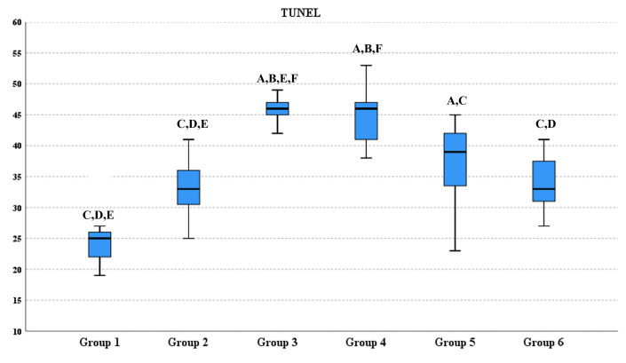
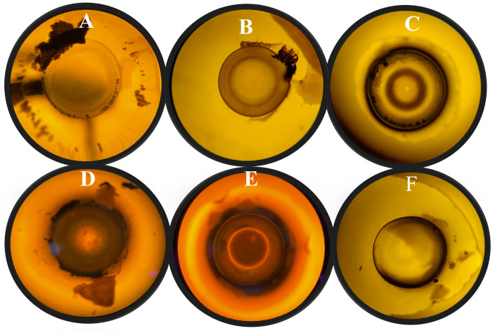
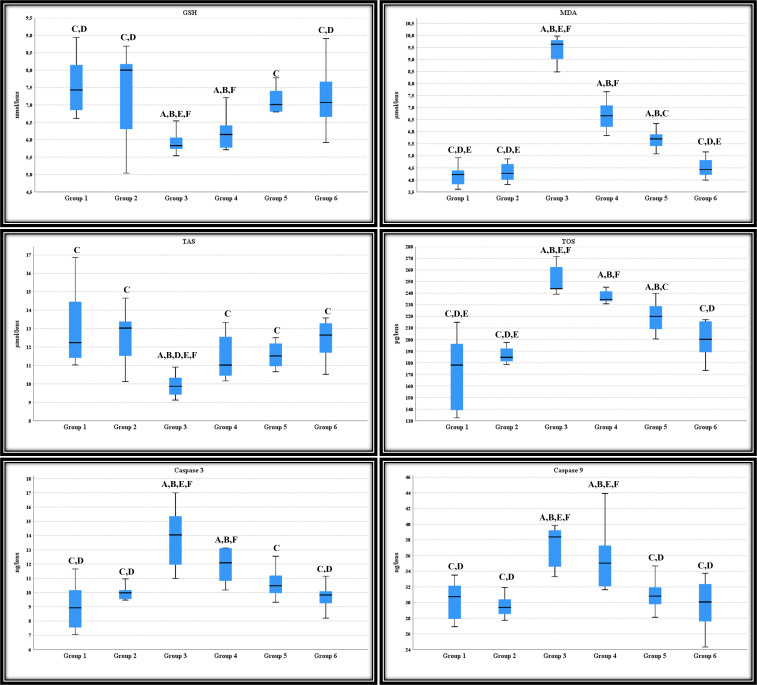
Methods: Within the scope of the study, 150 specific pathogen free (SPF) fertilized eggs were used. Eggs were divided into 6 groups as control (group 1), corn oil (group 2), hydrocortisone hemisuccinate sodium (HC) (group 3), low dose chrysin (group 4), medium dose chrysin (group 5) and high dose chrysin (group 6). On the 15th day of incubation, Chrysin and HC were applicated to the air sac of the eggs with Hamilton and/or insulin injector. On day 17, the chick embryos were removed from the eggs and the bulbus oculi of the embryos were dissected. Lenses of 9 embryos were used for morpholigical cataract grading in each group, lens of 8 embryos for biochemical analysis and intact eyes of 7 embryos for histological evaluation (TUNEL method).
Results: No opacity was observed in any of the lenses in Group 1 and 2. Cataract was observed in all lenses in Group 3. The mean opacity grades in group 3 were statistically significantly higher when compared to group 1 and 2 (p<0.05). The difference between group 6 and group 3 was statistically significant (p<0.05). GSH and TAS levels in the lenses were statistically significantly decreased compared to the control group due to HC application (p<0.05). It was determined that the decreased GSH and TAS levels in the lenses increased in relation to the Chrysin application doses. The increased levels of MDA, TOS, caspase 3 and caspase 9 in the HC group decreased significantly depending to the chrysin doses (p<0.05). In addition, while the rate of apoptotic cells determined by the TUNEL method was statistically significantly higher in the HC administered group than in the control group (p<0.05), it was statistically significantly decreased in the chrysin-administered groups, in relation to the dose of chrysin (p<0.05).
Conclusions: We think that anti-cataract effect of crhysin may be due to the antioxidant and antiapoptotic properties of chrysin. However, more research is needed to clarify the anti-cataract effects of chrysin.
期刊介绍:
Molecular Vision is a peer-reviewed journal dedicated to the dissemination of research results in molecular biology, cell biology, and the genetics of the visual system (ocular and cortical).
Molecular Vision publishes articles presenting original research that has not previously been published and comprehensive articles reviewing the current status of a particular field or topic. Submissions to Molecular Vision are subjected to rigorous peer review. Molecular Vision does NOT publish preprints.
For authors, Molecular Vision provides a rapid means of communicating important results. Access to Molecular Vision is free and unrestricted, allowing the widest possible audience for your article. Digital publishing allows you to use color images freely (and without fees). Additionally, you may publish animations, sounds, or other supplementary information that clarifies or supports your article. Each of the authors of an article may also list an electronic mail address (which will be updated upon request) to give interested readers easy access to authors.
 分享
分享


 求助内容:
求助内容: 应助结果提醒方式:
 扫码关注我们
