S A Golyshev, E P Kazakov, I I Kireev, D G Reunov, I V Malyshev
{"title":"细胞生物学中的软 X 射线显微镜:现状、贡献和前景。","authors":"S A Golyshev, E P Kazakov, I I Kireev, D G Reunov, I V Malyshev","doi":"10.32607/actanaturae.26551","DOIUrl":null,"url":null,"abstract":"<p><p>The recent advances achieved in microscopy technology have led to a significant breakthrough in biological research. Super-resolution fluorescent microscopy now allows us to visualize subcellular structures down to the pin-pointing of the single molecules in them, while modern electron microscopy has opened new possibilities in the study of protein complexes in their native, intracellular environment at near-atomic resolution. Nonetheless, both fluorescent and electron microscopy have remained beset by their principal shortcomings: the reliance on labeling procedures and severe sample volume limitations, respectively. Soft X-ray microscopy is a candidate method that can compensate for the shortcomings of both technologies by making possible observation of the entirety of the cellular interior without chemical fixation and labeling with an isotropic resolution of 40-70 nm. This will thus bridge the resolution gap between light and electron microscopy (although this gap is being narrowed, it still exists) and resolve the issue of compatibility with the former, and possibly in the near future, the latter methods. This review aims to assess the current state of soft X-ray microscopy and its impact on our understanding of the subcellular organization. It also attempts to look into the future of X-ray microscopy, particularly as relates to its seamless integration into the cell biology toolkit.</p>","PeriodicalId":6989,"journal":{"name":"Acta Naturae","volume":"15 4","pages":"32-43"},"PeriodicalIF":2.0000,"publicationDate":"2023-10-01","publicationTypes":"Journal Article","fieldsOfStudy":null,"isOpenAccess":false,"openAccessPdf":"https://www.ncbi.nlm.nih.gov/pmc/articles/PMC10790358/pdf/","citationCount":"0","resultStr":"{\"title\":\"Soft X-ray Microscopy in Cell Biology: Current Status, Contributions and Prospects.\",\"authors\":\"S A Golyshev, E P Kazakov, I I Kireev, D G Reunov, I V Malyshev\",\"doi\":\"10.32607/actanaturae.26551\",\"DOIUrl\":null,\"url\":null,\"abstract\":\"<p><p>The recent advances achieved in microscopy technology have led to a significant breakthrough in biological research. Super-resolution fluorescent microscopy now allows us to visualize subcellular structures down to the pin-pointing of the single molecules in them, while modern electron microscopy has opened new possibilities in the study of protein complexes in their native, intracellular environment at near-atomic resolution. Nonetheless, both fluorescent and electron microscopy have remained beset by their principal shortcomings: the reliance on labeling procedures and severe sample volume limitations, respectively. Soft X-ray microscopy is a candidate method that can compensate for the shortcomings of both technologies by making possible observation of the entirety of the cellular interior without chemical fixation and labeling with an isotropic resolution of 40-70 nm. This will thus bridge the resolution gap between light and electron microscopy (although this gap is being narrowed, it still exists) and resolve the issue of compatibility with the former, and possibly in the near future, the latter methods. This review aims to assess the current state of soft X-ray microscopy and its impact on our understanding of the subcellular organization. It also attempts to look into the future of X-ray microscopy, particularly as relates to its seamless integration into the cell biology toolkit.</p>\",\"PeriodicalId\":6989,\"journal\":{\"name\":\"Acta Naturae\",\"volume\":\"15 4\",\"pages\":\"32-43\"},\"PeriodicalIF\":2.0000,\"publicationDate\":\"2023-10-01\",\"publicationTypes\":\"Journal Article\",\"fieldsOfStudy\":null,\"isOpenAccess\":false,\"openAccessPdf\":\"https://www.ncbi.nlm.nih.gov/pmc/articles/PMC10790358/pdf/\",\"citationCount\":\"0\",\"resultStr\":null,\"platform\":\"Semanticscholar\",\"paperid\":null,\"PeriodicalName\":\"Acta Naturae\",\"FirstCategoryId\":\"99\",\"ListUrlMain\":\"https://doi.org/10.32607/actanaturae.26551\",\"RegionNum\":4,\"RegionCategory\":\"生物学\",\"ArticlePicture\":[],\"TitleCN\":null,\"AbstractTextCN\":null,\"PMCID\":null,\"EPubDate\":\"\",\"PubModel\":\"\",\"JCR\":\"Q4\",\"JCRName\":\"CELL BIOLOGY\",\"Score\":null,\"Total\":0}","platform":"Semanticscholar","paperid":null,"PeriodicalName":"Acta Naturae","FirstCategoryId":"99","ListUrlMain":"https://doi.org/10.32607/actanaturae.26551","RegionNum":4,"RegionCategory":"生物学","ArticlePicture":[],"TitleCN":null,"AbstractTextCN":null,"PMCID":null,"EPubDate":"","PubModel":"","JCR":"Q4","JCRName":"CELL BIOLOGY","Score":null,"Total":0}
引用次数: 0
摘要
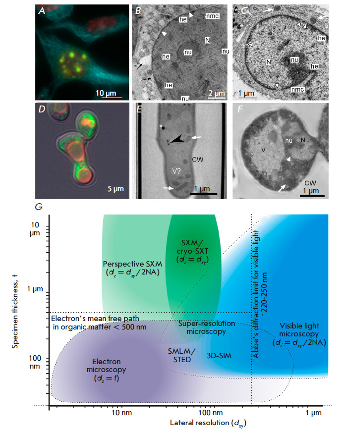
显微镜技术的最新进展为生物研究带来了重大突破。现在,超分辨率荧光显微镜使我们能够观察到亚细胞结构,甚至能够精确定位其中的单个分子,而现代电子显微镜则为研究原生细胞内环境中的蛋白质复合物提供了近乎原子分辨率的新可能性。尽管如此,荧光显微镜和电子显微镜仍然存在主要缺陷:分别依赖于标记程序和严重的样品体积限制。软 X 射线显微镜是一种可弥补这两种技术不足的候选方法,它无需化学固定和标记即可观察到整个细胞内部,各向同性分辨率可达 40-70 纳米。这将弥补光学显微镜和电子显微镜之间的分辨率差距(虽然这一差距正在缩小,但仍然存在),并解决与前者的兼容性问题,在不久的将来还可能解决与后者的兼容性问题。本综述旨在评估软 X 射线显微镜的现状及其对我们了解亚细胞组织的影响。它还试图展望 X 射线显微镜的未来,特别是它与细胞生物学工具包的无缝整合。
Soft X-ray Microscopy in Cell Biology: Current Status, Contributions and Prospects.
The recent advances achieved in microscopy technology have led to a significant breakthrough in biological research. Super-resolution fluorescent microscopy now allows us to visualize subcellular structures down to the pin-pointing of the single molecules in them, while modern electron microscopy has opened new possibilities in the study of protein complexes in their native, intracellular environment at near-atomic resolution. Nonetheless, both fluorescent and electron microscopy have remained beset by their principal shortcomings: the reliance on labeling procedures and severe sample volume limitations, respectively. Soft X-ray microscopy is a candidate method that can compensate for the shortcomings of both technologies by making possible observation of the entirety of the cellular interior without chemical fixation and labeling with an isotropic resolution of 40-70 nm. This will thus bridge the resolution gap between light and electron microscopy (although this gap is being narrowed, it still exists) and resolve the issue of compatibility with the former, and possibly in the near future, the latter methods. This review aims to assess the current state of soft X-ray microscopy and its impact on our understanding of the subcellular organization. It also attempts to look into the future of X-ray microscopy, particularly as relates to its seamless integration into the cell biology toolkit.
期刊介绍:
Acta Naturae is an international journal on life sciences based in Moscow, Russia.
Our goal is to present scientific work and discovery in molecular biology, biochemistry, biomedical disciplines and biotechnology. These fields represent the most important priorities for the research and engineering development both in Russia and worldwide. Acta Naturae is also a periodical for those who are curious in various aspects of biotechnological business, innovations in pharmaceutical areas, intellectual property protection and social consequences of scientific progress. The journal publishes analytical industrial surveys focused on the development of different spheres of modern life science and technology.
Being a radically new and totally unique journal in Russia, Acta Naturae is useful to both representatives of fundamental research and experts in applied sciences.


 分享
分享
 求助内容:
求助内容: 应助结果提醒方式:
 扫码关注我们
