Lu Lu Xie, Ying Gong, Kui Ran Dong, Chun Shen, Bo Duan, Rui Dong
{"title":"应用机器学习和深度高效网络从增强计算机断层扫描图像中区分新生儿肾上腺血肿和神经母细胞瘤","authors":"Lu Lu Xie, Ying Gong, Kui Ran Dong, Chun Shen, Bo Duan, Rui Dong","doi":"10.14740/wjon1744","DOIUrl":null,"url":null,"abstract":"<p><strong>Background: </strong>The aim of the study was to employ a combination of radiomic indicators based on computed tomography (CT) imaging and machine learning (ML), along with deep learning (DL), to differentiate between adrenal hematoma and adrenal neuroblastoma in neonates.</p><p><strong>Methods: </strong>A total of 76 neonates were included in this retrospective study (40 with neuroblastomas and 36 with adrenal hematomas) who underwent CT and divided into a training group (n = 38) and a testing group (n = 38). The regions of interest (ROIs) were segmented by two radiologists to extract radiomics features using Pyradiomics package. ML classifications were done using support vector machine (SVM), AdaBoost, Extra Trees, gradient boosting, multi-layer perceptron (MLP), and random forest (RF). EfficientNets was employed and classified, based on radiometrics. The area under curve (AUC) of the receiver operating characteristic (ROC) was calculated to assess the performance of each model.</p><p><strong>Results: </strong>Among all features, the least absolute shrinkage and selection operator (LASSO) logistic regression selected nine features. These radiomics features were used to construct radiomics model. In the training cohort, the AUCs of SVM, MLP and Extra Trees models were 0.967, 0.969 and 1.000, respectively. The corresponding AUCs of the test cohort were 0.985, 0.971 and 0.958, respectively. In the classification task, the AUC of the DL framework was 0.987.</p><p><strong>Conclusion: </strong>ML decision classifiers and DL framework constructed from CT-based radiomics features offered a non-invasive method to differentiate neonatal adrenal hematoma from neuroblastoma and performed better than the clinical experts.</p>","PeriodicalId":46797,"journal":{"name":"World Journal of Oncology","volume":"15 1","pages":"81-89"},"PeriodicalIF":2.2000,"publicationDate":"2024-02-01","publicationTypes":"Journal Article","fieldsOfStudy":null,"isOpenAccess":false,"openAccessPdf":"https://www.ncbi.nlm.nih.gov/pmc/articles/PMC10807921/pdf/","citationCount":"0","resultStr":"{\"title\":\"Application of Machine Learning and Deep EfficientNets in Distinguishing Neonatal Adrenal Hematomas From Neuroblastoma in Enhanced Computed Tomography Images.\",\"authors\":\"Lu Lu Xie, Ying Gong, Kui Ran Dong, Chun Shen, Bo Duan, Rui Dong\",\"doi\":\"10.14740/wjon1744\",\"DOIUrl\":null,\"url\":null,\"abstract\":\"<p><strong>Background: </strong>The aim of the study was to employ a combination of radiomic indicators based on computed tomography (CT) imaging and machine learning (ML), along with deep learning (DL), to differentiate between adrenal hematoma and adrenal neuroblastoma in neonates.</p><p><strong>Methods: </strong>A total of 76 neonates were included in this retrospective study (40 with neuroblastomas and 36 with adrenal hematomas) who underwent CT and divided into a training group (n = 38) and a testing group (n = 38). The regions of interest (ROIs) were segmented by two radiologists to extract radiomics features using Pyradiomics package. ML classifications were done using support vector machine (SVM), AdaBoost, Extra Trees, gradient boosting, multi-layer perceptron (MLP), and random forest (RF). EfficientNets was employed and classified, based on radiometrics. The area under curve (AUC) of the receiver operating characteristic (ROC) was calculated to assess the performance of each model.</p><p><strong>Results: </strong>Among all features, the least absolute shrinkage and selection operator (LASSO) logistic regression selected nine features. These radiomics features were used to construct radiomics model. In the training cohort, the AUCs of SVM, MLP and Extra Trees models were 0.967, 0.969 and 1.000, respectively. The corresponding AUCs of the test cohort were 0.985, 0.971 and 0.958, respectively. In the classification task, the AUC of the DL framework was 0.987.</p><p><strong>Conclusion: </strong>ML decision classifiers and DL framework constructed from CT-based radiomics features offered a non-invasive method to differentiate neonatal adrenal hematoma from neuroblastoma and performed better than the clinical experts.</p>\",\"PeriodicalId\":46797,\"journal\":{\"name\":\"World Journal of Oncology\",\"volume\":\"15 1\",\"pages\":\"81-89\"},\"PeriodicalIF\":2.2000,\"publicationDate\":\"2024-02-01\",\"publicationTypes\":\"Journal Article\",\"fieldsOfStudy\":null,\"isOpenAccess\":false,\"openAccessPdf\":\"https://www.ncbi.nlm.nih.gov/pmc/articles/PMC10807921/pdf/\",\"citationCount\":\"0\",\"resultStr\":null,\"platform\":\"Semanticscholar\",\"paperid\":null,\"PeriodicalName\":\"World Journal of Oncology\",\"FirstCategoryId\":\"1085\",\"ListUrlMain\":\"https://doi.org/10.14740/wjon1744\",\"RegionNum\":0,\"RegionCategory\":null,\"ArticlePicture\":[],\"TitleCN\":null,\"AbstractTextCN\":null,\"PMCID\":null,\"EPubDate\":\"2024/1/20 0:00:00\",\"PubModel\":\"Epub\",\"JCR\":\"Q3\",\"JCRName\":\"ONCOLOGY\",\"Score\":null,\"Total\":0}","platform":"Semanticscholar","paperid":null,"PeriodicalName":"World Journal of Oncology","FirstCategoryId":"1085","ListUrlMain":"https://doi.org/10.14740/wjon1744","RegionNum":0,"RegionCategory":null,"ArticlePicture":[],"TitleCN":null,"AbstractTextCN":null,"PMCID":null,"EPubDate":"2024/1/20 0:00:00","PubModel":"Epub","JCR":"Q3","JCRName":"ONCOLOGY","Score":null,"Total":0}
Application of Machine Learning and Deep EfficientNets in Distinguishing Neonatal Adrenal Hematomas From Neuroblastoma in Enhanced Computed Tomography Images.
Background: The aim of the study was to employ a combination of radiomic indicators based on computed tomography (CT) imaging and machine learning (ML), along with deep learning (DL), to differentiate between adrenal hematoma and adrenal neuroblastoma in neonates.
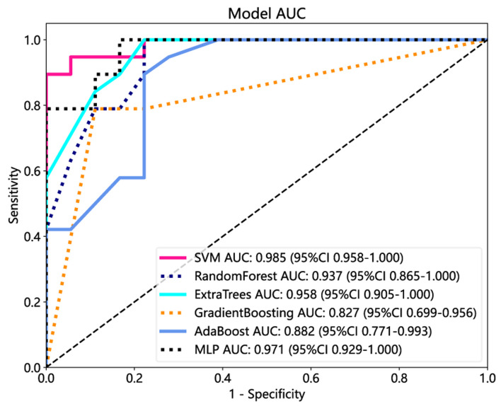
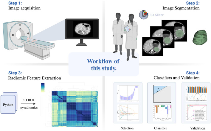
Methods: A total of 76 neonates were included in this retrospective study (40 with neuroblastomas and 36 with adrenal hematomas) who underwent CT and divided into a training group (n = 38) and a testing group (n = 38). The regions of interest (ROIs) were segmented by two radiologists to extract radiomics features using Pyradiomics package. ML classifications were done using support vector machine (SVM), AdaBoost, Extra Trees, gradient boosting, multi-layer perceptron (MLP), and random forest (RF). EfficientNets was employed and classified, based on radiometrics. The area under curve (AUC) of the receiver operating characteristic (ROC) was calculated to assess the performance of each model.
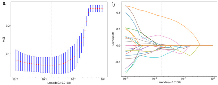
Results: Among all features, the least absolute shrinkage and selection operator (LASSO) logistic regression selected nine features. These radiomics features were used to construct radiomics model. In the training cohort, the AUCs of SVM, MLP and Extra Trees models were 0.967, 0.969 and 1.000, respectively. The corresponding AUCs of the test cohort were 0.985, 0.971 and 0.958, respectively. In the classification task, the AUC of the DL framework was 0.987.
Conclusion: ML decision classifiers and DL framework constructed from CT-based radiomics features offered a non-invasive method to differentiate neonatal adrenal hematoma from neuroblastoma and performed better than the clinical experts.
期刊介绍:
World Journal of Oncology, bimonthly, publishes original contributions describing basic research and clinical investigation of cancer, on the cellular, molecular, prevention, diagnosis, therapy and prognosis aspects. The submissions can be basic research or clinical investigation oriented. This journal welcomes those submissions focused on the clinical trials of new treatment modalities for cancer, and those submissions focused on molecular or cellular research of the oncology pathogenesis. Case reports submitted for consideration of publication should explore either a novel genomic event/description or a new safety signal from an oncolytic agent. The areas of interested manuscripts are these disciplines: tumor immunology and immunotherapy; cancer molecular pharmacology and chemotherapy; drug sensitivity and resistance; cancer epidemiology; clinical trials; cancer pathology; radiobiology and radiation oncology; solid tumor oncology; hematological malignancies; surgical oncology; pediatric oncology; molecular oncology and cancer genes; gene therapy; cancer endocrinology; cancer metastasis; prevention and diagnosis of cancer; other cancer related subjects. The types of manuscripts accepted are original article, review, editorial, short communication, case report, letter to the editor, book review.


 分享
分享
 求助内容:
求助内容: 应助结果提醒方式:
 扫码关注我们
