Matheus Junger do Brasil, Clara Villela Castello Branco, Naomi Cappelli, Joice Bento da Silva, Rosaura Leite Rodrigues, Maria Eduarda Dos Santos Lopes Fernandes
{"title":"猫狗浅表淋巴腺切除术图解技术:初步研究。","authors":"Matheus Junger do Brasil, Clara Villela Castello Branco, Naomi Cappelli, Joice Bento da Silva, Rosaura Leite Rodrigues, Maria Eduarda Dos Santos Lopes Fernandes","doi":"10.29374/2527-2179.bjvm004823","DOIUrl":null,"url":null,"abstract":"<p><p>Superficial lymphadenectomy is an easy-to-perform and cost-effective routine technique. Despite its simplicity, it remains underutilized in veterinary medicine, with most practitioners being oncological surgeons. This study aims to enhance accessibility to the surgical procedure by providing anatomical representations of superficial lymphadenectomy in the carcasses of dogs and cats. A preliminary study involving two canines and two felines was conducted, with each group comprising a dog and a cat. Group A was designated to superficial lymphadenectomy techniques to create an illustrated step-by-step procedure, while group B underwent anatomical dissection to expose lymph nodes and their adnexa. The approach to superficial lymph nodes in dogs and cats is simple, allowing for the demonstration of superficial lymphadenectomy techniques in the corpses of dogs and cats without complications. This includes the dissection and presentation of anatomical structures adjacent to the lymph nodes. In conclusion, the techniques applied to subjects in groups A and B proved effective, successfully demonstrating and excising all superficial lymph nodes in the corpses of dogs and cats. These findings suggest that the developed set of techniques developed for lymph node excision holds promise for safe and effective application in live animals.</p>","PeriodicalId":72458,"journal":{"name":"Brazilian journal of veterinary medicine","volume":"46 ","pages":"e004823"},"PeriodicalIF":0.0000,"publicationDate":"2024-01-17","publicationTypes":"Journal Article","fieldsOfStudy":null,"isOpenAccess":false,"openAccessPdf":"https://www.ncbi.nlm.nih.gov/pmc/articles/PMC10811725/pdf/","citationCount":"0","resultStr":"{\"title\":\"Illustrated technique of superficial lymphadenectomy of dogs and cats: preliminary study.\",\"authors\":\"Matheus Junger do Brasil, Clara Villela Castello Branco, Naomi Cappelli, Joice Bento da Silva, Rosaura Leite Rodrigues, Maria Eduarda Dos Santos Lopes Fernandes\",\"doi\":\"10.29374/2527-2179.bjvm004823\",\"DOIUrl\":null,\"url\":null,\"abstract\":\"<p><p>Superficial lymphadenectomy is an easy-to-perform and cost-effective routine technique. Despite its simplicity, it remains underutilized in veterinary medicine, with most practitioners being oncological surgeons. This study aims to enhance accessibility to the surgical procedure by providing anatomical representations of superficial lymphadenectomy in the carcasses of dogs and cats. A preliminary study involving two canines and two felines was conducted, with each group comprising a dog and a cat. Group A was designated to superficial lymphadenectomy techniques to create an illustrated step-by-step procedure, while group B underwent anatomical dissection to expose lymph nodes and their adnexa. The approach to superficial lymph nodes in dogs and cats is simple, allowing for the demonstration of superficial lymphadenectomy techniques in the corpses of dogs and cats without complications. This includes the dissection and presentation of anatomical structures adjacent to the lymph nodes. In conclusion, the techniques applied to subjects in groups A and B proved effective, successfully demonstrating and excising all superficial lymph nodes in the corpses of dogs and cats. These findings suggest that the developed set of techniques developed for lymph node excision holds promise for safe and effective application in live animals.</p>\",\"PeriodicalId\":72458,\"journal\":{\"name\":\"Brazilian journal of veterinary medicine\",\"volume\":\"46 \",\"pages\":\"e004823\"},\"PeriodicalIF\":0.0000,\"publicationDate\":\"2024-01-17\",\"publicationTypes\":\"Journal Article\",\"fieldsOfStudy\":null,\"isOpenAccess\":false,\"openAccessPdf\":\"https://www.ncbi.nlm.nih.gov/pmc/articles/PMC10811725/pdf/\",\"citationCount\":\"0\",\"resultStr\":null,\"platform\":\"Semanticscholar\",\"paperid\":null,\"PeriodicalName\":\"Brazilian journal of veterinary medicine\",\"FirstCategoryId\":\"1085\",\"ListUrlMain\":\"https://doi.org/10.29374/2527-2179.bjvm004823\",\"RegionNum\":0,\"RegionCategory\":null,\"ArticlePicture\":[],\"TitleCN\":null,\"AbstractTextCN\":null,\"PMCID\":null,\"EPubDate\":\"2024/1/1 0:00:00\",\"PubModel\":\"eCollection\",\"JCR\":\"\",\"JCRName\":\"\",\"Score\":null,\"Total\":0}","platform":"Semanticscholar","paperid":null,"PeriodicalName":"Brazilian journal of veterinary medicine","FirstCategoryId":"1085","ListUrlMain":"https://doi.org/10.29374/2527-2179.bjvm004823","RegionNum":0,"RegionCategory":null,"ArticlePicture":[],"TitleCN":null,"AbstractTextCN":null,"PMCID":null,"EPubDate":"2024/1/1 0:00:00","PubModel":"eCollection","JCR":"","JCRName":"","Score":null,"Total":0}
引用次数: 0
摘要
浅表淋巴结切除术是一种易于操作、成本效益高的常规技术。尽管操作简单,但在兽医学中仍未得到充分利用,大多数从业者都是肿瘤外科医生。本研究旨在通过提供猫狗尸体浅表淋巴腺切除术的解剖图,提高手术的可及性。我们进行了一项涉及两只犬和两只猫的初步研究,每组包括一只犬和一只猫。A 组指定浅表淋巴结切除技术,以创建一个图文并茂的步骤,而 B 组则进行解剖解剖,以暴露淋巴结及其附件。处理猫狗浅表淋巴结的方法很简单,可以在没有并发症的情况下展示猫狗尸体浅表淋巴结切除技术。这包括解剖和展示淋巴结附近的解剖结构。总之,在 A 组和 B 组受试者身上应用的技术证明是有效的,成功地展示并切除了猫狗尸体上的所有浅表淋巴结。这些研究结果表明,所开发的这套淋巴结切除技术有望在活体动物身上安全有效地应用。
Illustrated technique of superficial lymphadenectomy of dogs and cats: preliminary study.
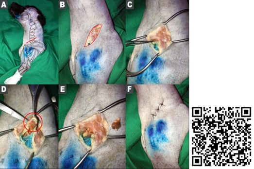
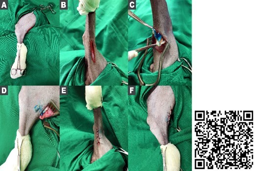
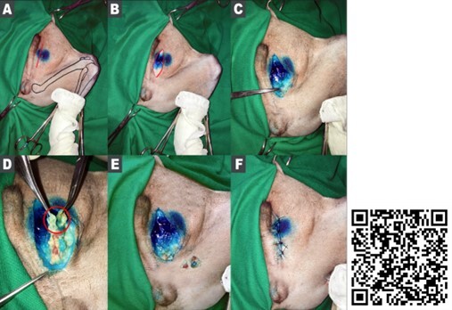
Superficial lymphadenectomy is an easy-to-perform and cost-effective routine technique. Despite its simplicity, it remains underutilized in veterinary medicine, with most practitioners being oncological surgeons. This study aims to enhance accessibility to the surgical procedure by providing anatomical representations of superficial lymphadenectomy in the carcasses of dogs and cats. A preliminary study involving two canines and two felines was conducted, with each group comprising a dog and a cat. Group A was designated to superficial lymphadenectomy techniques to create an illustrated step-by-step procedure, while group B underwent anatomical dissection to expose lymph nodes and their adnexa. The approach to superficial lymph nodes in dogs and cats is simple, allowing for the demonstration of superficial lymphadenectomy techniques in the corpses of dogs and cats without complications. This includes the dissection and presentation of anatomical structures adjacent to the lymph nodes. In conclusion, the techniques applied to subjects in groups A and B proved effective, successfully demonstrating and excising all superficial lymph nodes in the corpses of dogs and cats. These findings suggest that the developed set of techniques developed for lymph node excision holds promise for safe and effective application in live animals.


 分享
分享

 求助内容:
求助内容: 应助结果提醒方式:
 扫码关注我们
