{"title":"与阿糖胞苷相关的迟发性全身红斑的临床特征","authors":"He Jiang, Jun Lu, Mei Wang","doi":"10.1002/cdt3.117","DOIUrl":null,"url":null,"abstract":"<p>Cytarabine is one of the most used drugs for the treatment of hematological malignancies such as leukemia and non-Hodgkin's lymphoma. In cells, cytarabine is activated into ara-CTP, which can replace deoxycytidine triphosphate (dCTP) and become incorporated into the DNA of proliferating cells. Thus, it can block DNA synthesis, resulting in proliferation arrest and cell death.<span><sup>1</sup></span> High doses of cytarabine usually induce dermatological toxicity commonly reported as morbilliform eruptions, acral erythema, neutrophilic eccrine hidradenitis, vasculitis, toxic epidermal necrolysis, and eccrine squamous syringome taplasia.<span><sup>2-5</sup></span> Of these toxicity effects, violaceous erythema is especially rare.<span><sup>6, 7</sup></span> Herein, we present a case of delayed generalized violaceous erythema associated with cytarabine and discuss its necessary treatment.</p><p>An 11-year-old boy came to the hospital complaining of an ache in the lower limb. Bone marrow puncture results confirm that he has acute lymphoblastic leukemia. He received a chemotherapy with cyclophosphamide, cytarabine, and 6-mercaptopurine (according to the Chinese Children Cancer Group [CCCG]-Acute lymphoblastic leukemia [ALL]-2015) for induction chemotherapy. The treatment course lasted 7 days, with cytarabine (100 mg/m<sup>2</sup>) and 6-mercaptopurine (60 mg/m<sup>2</sup>) given daily. Cyclophosphamide (1 g/m<sup>2</sup>) was given on the first day. On the seventh day of chemotherapy, he developed a high fever (39.8°C). The next day, a florid, diffuse, nonpruritic erythematous macule eruption appeared on his face, ears, and scalp. Antihistamines have no effect on the erythematous macule. Over the next 24 h, erythematous macules spread over his neck, arms, chest, abdomen, and back (Figure 1A). The percentage of eosinophils (6%; reference value, 0–5%) increased transiently on the eighth day of chemotherapy. On the ninth day of chemotherapy, erythematous macules coalesced into purpuric plaques and spread throughout the body (Figure 1B). The color of the erythema turned to bright red as well as a growing facial edema was observed. Histopathological examination of skin biopsy tissue demonstrated neutrophilic, lymphocytic infiltration and erythrocytic extravasation (Figure 2). C-reactive protein (CRP) level (56.7 mg/L; reference value, 0–8 mg/L) and D-dimer (2860 µg/L; reference value, 0–550 µg/L) were high throughout the period of erythema. Meanwhile, anti-infective therapy showed no effect on the progression of erythema or the reduction of CRP. On the 12th day of initial cytarabine exposure, dry desquamation was noted (Figure 1C), with complaints of slight pruritus. Then, the eruption faded away and disappeared on the 17th day after initial exposure to cytarabine (Figure 1D). However, in the later stage of erythema, the patient developed a liver injury and lasted for 13 days. After 28 days of initial exposure to cytarabine, the patient recovered. Subsequently, he received a same chemotherapy and the erythema did not reappear.</p><p>The cytarabine-associated erythema had the following characteristics. (1) The initial rash was accompanied by a high fever, and the rash could rapidly expand all over the body within one or 2 days. (2) After the eruption, there was a facial edema, including the eyelids. (3) Elevated eosinophilia occurred at the most severe erythema period. (4) During the erythema, there was an increase in CRP without microbial infection. (5) There was a dramatic increase in platelet-independent D-dimer during the eruption. (6) It is self-limiting and may occur after the cytarabine treatment. (7) Patients may develop organ damage after the erythema.</p><p>Here we reported a kind of specific skin toxicity reaction of cytarabine. We have presented in detail with the characteristic morphology, distribution characteristics, and the timeline of the cytarabine-associated erythema. It will be helpful for clinical diagnosis and treatment. Confidence in its benign nature will prevent unnecessary intervention or cessation of chemotherapy. More attention should be paid to the damage of internal organs while treating with the erythema.</p><p>He Jiang and Mei Wang reviewed the literature and wrote the manuscript. Mei Wang and Jun Lu supervised and revised the manuscript. All authors read and approved the final manuscript.</p><p>The authors declare no conflict of interest.</p><p>The Committee on Human Research at the Children's Hospital of Soochow University reviewed and approved this study (2023CS201) and informed consents have been properly documented.</p>","PeriodicalId":32096,"journal":{"name":"Chronic Diseases and Translational Medicine","volume":"10 1","pages":"78-80"},"PeriodicalIF":0.0000,"publicationDate":"2024-01-27","publicationTypes":"Journal Article","fieldsOfStudy":null,"isOpenAccess":false,"openAccessPdf":"https://onlinelibrary.wiley.com/doi/epdf/10.1002/cdt3.117","citationCount":"0","resultStr":"{\"title\":\"Clinical characteristics of delayed generalized erythema associated with cytarabine\",\"authors\":\"He Jiang, Jun Lu, Mei Wang\",\"doi\":\"10.1002/cdt3.117\",\"DOIUrl\":null,\"url\":null,\"abstract\":\"<p>Cytarabine is one of the most used drugs for the treatment of hematological malignancies such as leukemia and non-Hodgkin's lymphoma. In cells, cytarabine is activated into ara-CTP, which can replace deoxycytidine triphosphate (dCTP) and become incorporated into the DNA of proliferating cells. Thus, it can block DNA synthesis, resulting in proliferation arrest and cell death.<span><sup>1</sup></span> High doses of cytarabine usually induce dermatological toxicity commonly reported as morbilliform eruptions, acral erythema, neutrophilic eccrine hidradenitis, vasculitis, toxic epidermal necrolysis, and eccrine squamous syringome taplasia.<span><sup>2-5</sup></span> Of these toxicity effects, violaceous erythema is especially rare.<span><sup>6, 7</sup></span> Herein, we present a case of delayed generalized violaceous erythema associated with cytarabine and discuss its necessary treatment.</p><p>An 11-year-old boy came to the hospital complaining of an ache in the lower limb. Bone marrow puncture results confirm that he has acute lymphoblastic leukemia. He received a chemotherapy with cyclophosphamide, cytarabine, and 6-mercaptopurine (according to the Chinese Children Cancer Group [CCCG]-Acute lymphoblastic leukemia [ALL]-2015) for induction chemotherapy. The treatment course lasted 7 days, with cytarabine (100 mg/m<sup>2</sup>) and 6-mercaptopurine (60 mg/m<sup>2</sup>) given daily. Cyclophosphamide (1 g/m<sup>2</sup>) was given on the first day. On the seventh day of chemotherapy, he developed a high fever (39.8°C). The next day, a florid, diffuse, nonpruritic erythematous macule eruption appeared on his face, ears, and scalp. Antihistamines have no effect on the erythematous macule. Over the next 24 h, erythematous macules spread over his neck, arms, chest, abdomen, and back (Figure 1A). The percentage of eosinophils (6%; reference value, 0–5%) increased transiently on the eighth day of chemotherapy. On the ninth day of chemotherapy, erythematous macules coalesced into purpuric plaques and spread throughout the body (Figure 1B). The color of the erythema turned to bright red as well as a growing facial edema was observed. Histopathological examination of skin biopsy tissue demonstrated neutrophilic, lymphocytic infiltration and erythrocytic extravasation (Figure 2). C-reactive protein (CRP) level (56.7 mg/L; reference value, 0–8 mg/L) and D-dimer (2860 µg/L; reference value, 0–550 µg/L) were high throughout the period of erythema. Meanwhile, anti-infective therapy showed no effect on the progression of erythema or the reduction of CRP. On the 12th day of initial cytarabine exposure, dry desquamation was noted (Figure 1C), with complaints of slight pruritus. Then, the eruption faded away and disappeared on the 17th day after initial exposure to cytarabine (Figure 1D). However, in the later stage of erythema, the patient developed a liver injury and lasted for 13 days. After 28 days of initial exposure to cytarabine, the patient recovered. Subsequently, he received a same chemotherapy and the erythema did not reappear.</p><p>The cytarabine-associated erythema had the following characteristics. (1) The initial rash was accompanied by a high fever, and the rash could rapidly expand all over the body within one or 2 days. (2) After the eruption, there was a facial edema, including the eyelids. (3) Elevated eosinophilia occurred at the most severe erythema period. (4) During the erythema, there was an increase in CRP without microbial infection. (5) There was a dramatic increase in platelet-independent D-dimer during the eruption. (6) It is self-limiting and may occur after the cytarabine treatment. (7) Patients may develop organ damage after the erythema.</p><p>Here we reported a kind of specific skin toxicity reaction of cytarabine. We have presented in detail with the characteristic morphology, distribution characteristics, and the timeline of the cytarabine-associated erythema. It will be helpful for clinical diagnosis and treatment. Confidence in its benign nature will prevent unnecessary intervention or cessation of chemotherapy. More attention should be paid to the damage of internal organs while treating with the erythema.</p><p>He Jiang and Mei Wang reviewed the literature and wrote the manuscript. Mei Wang and Jun Lu supervised and revised the manuscript. All authors read and approved the final manuscript.</p><p>The authors declare no conflict of interest.</p><p>The Committee on Human Research at the Children's Hospital of Soochow University reviewed and approved this study (2023CS201) and informed consents have been properly documented.</p>\",\"PeriodicalId\":32096,\"journal\":{\"name\":\"Chronic Diseases and Translational Medicine\",\"volume\":\"10 1\",\"pages\":\"78-80\"},\"PeriodicalIF\":0.0000,\"publicationDate\":\"2024-01-27\",\"publicationTypes\":\"Journal Article\",\"fieldsOfStudy\":null,\"isOpenAccess\":false,\"openAccessPdf\":\"https://onlinelibrary.wiley.com/doi/epdf/10.1002/cdt3.117\",\"citationCount\":\"0\",\"resultStr\":null,\"platform\":\"Semanticscholar\",\"paperid\":null,\"PeriodicalName\":\"Chronic Diseases and Translational Medicine\",\"FirstCategoryId\":\"3\",\"ListUrlMain\":\"https://onlinelibrary.wiley.com/doi/10.1002/cdt3.117\",\"RegionNum\":0,\"RegionCategory\":null,\"ArticlePicture\":[],\"TitleCN\":null,\"AbstractTextCN\":null,\"PMCID\":null,\"EPubDate\":\"\",\"PubModel\":\"\",\"JCR\":\"Q1\",\"JCRName\":\"Medicine\",\"Score\":null,\"Total\":0}","platform":"Semanticscholar","paperid":null,"PeriodicalName":"Chronic Diseases and Translational Medicine","FirstCategoryId":"3","ListUrlMain":"https://onlinelibrary.wiley.com/doi/10.1002/cdt3.117","RegionNum":0,"RegionCategory":null,"ArticlePicture":[],"TitleCN":null,"AbstractTextCN":null,"PMCID":null,"EPubDate":"","PubModel":"","JCR":"Q1","JCRName":"Medicine","Score":null,"Total":0}
Clinical characteristics of delayed generalized erythema associated with cytarabine
Cytarabine is one of the most used drugs for the treatment of hematological malignancies such as leukemia and non-Hodgkin's lymphoma. In cells, cytarabine is activated into ara-CTP, which can replace deoxycytidine triphosphate (dCTP) and become incorporated into the DNA of proliferating cells. Thus, it can block DNA synthesis, resulting in proliferation arrest and cell death.1 High doses of cytarabine usually induce dermatological toxicity commonly reported as morbilliform eruptions, acral erythema, neutrophilic eccrine hidradenitis, vasculitis, toxic epidermal necrolysis, and eccrine squamous syringome taplasia.2-5 Of these toxicity effects, violaceous erythema is especially rare.6, 7 Herein, we present a case of delayed generalized violaceous erythema associated with cytarabine and discuss its necessary treatment.
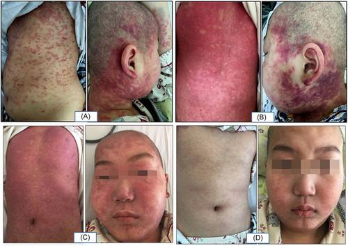
An 11-year-old boy came to the hospital complaining of an ache in the lower limb. Bone marrow puncture results confirm that he has acute lymphoblastic leukemia. He received a chemotherapy with cyclophosphamide, cytarabine, and 6-mercaptopurine (according to the Chinese Children Cancer Group [CCCG]-Acute lymphoblastic leukemia [ALL]-2015) for induction chemotherapy. The treatment course lasted 7 days, with cytarabine (100 mg/m2) and 6-mercaptopurine (60 mg/m2) given daily. Cyclophosphamide (1 g/m2) was given on the first day. On the seventh day of chemotherapy, he developed a high fever (39.8°C). The next day, a florid, diffuse, nonpruritic erythematous macule eruption appeared on his face, ears, and scalp. Antihistamines have no effect on the erythematous macule. Over the next 24 h, erythematous macules spread over his neck, arms, chest, abdomen, and back (Figure 1A). The percentage of eosinophils (6%; reference value, 0–5%) increased transiently on the eighth day of chemotherapy. On the ninth day of chemotherapy, erythematous macules coalesced into purpuric plaques and spread throughout the body (Figure 1B). The color of the erythema turned to bright red as well as a growing facial edema was observed. Histopathological examination of skin biopsy tissue demonstrated neutrophilic, lymphocytic infiltration and erythrocytic extravasation (Figure 2). C-reactive protein (CRP) level (56.7 mg/L; reference value, 0–8 mg/L) and D-dimer (2860 µg/L; reference value, 0–550 µg/L) were high throughout the period of erythema. Meanwhile, anti-infective therapy showed no effect on the progression of erythema or the reduction of CRP. On the 12th day of initial cytarabine exposure, dry desquamation was noted (Figure 1C), with complaints of slight pruritus. Then, the eruption faded away and disappeared on the 17th day after initial exposure to cytarabine (Figure 1D). However, in the later stage of erythema, the patient developed a liver injury and lasted for 13 days. After 28 days of initial exposure to cytarabine, the patient recovered. Subsequently, he received a same chemotherapy and the erythema did not reappear.
The cytarabine-associated erythema had the following characteristics. (1) The initial rash was accompanied by a high fever, and the rash could rapidly expand all over the body within one or 2 days. (2) After the eruption, there was a facial edema, including the eyelids. (3) Elevated eosinophilia occurred at the most severe erythema period. (4) During the erythema, there was an increase in CRP without microbial infection. (5) There was a dramatic increase in platelet-independent D-dimer during the eruption. (6) It is self-limiting and may occur after the cytarabine treatment. (7) Patients may develop organ damage after the erythema.
Here we reported a kind of specific skin toxicity reaction of cytarabine. We have presented in detail with the characteristic morphology, distribution characteristics, and the timeline of the cytarabine-associated erythema. It will be helpful for clinical diagnosis and treatment. Confidence in its benign nature will prevent unnecessary intervention or cessation of chemotherapy. More attention should be paid to the damage of internal organs while treating with the erythema.
He Jiang and Mei Wang reviewed the literature and wrote the manuscript. Mei Wang and Jun Lu supervised and revised the manuscript. All authors read and approved the final manuscript.
The authors declare no conflict of interest.
The Committee on Human Research at the Children's Hospital of Soochow University reviewed and approved this study (2023CS201) and informed consents have been properly documented.
期刊介绍:
This journal aims to promote progress from basic research to clinical practice and to provide a forum for communication among basic, translational, and clinical research practitioners and physicians from all relevant disciplines. Chronic diseases such as cardiovascular diseases, cancer, diabetes, stroke, chronic respiratory diseases (such as asthma and COPD), chronic kidney diseases, and related translational research. Topics of interest for Chronic Diseases and Translational Medicine include Research and commentary on models of chronic diseases with significant implications for disease diagnosis and treatment Investigative studies of human biology with an emphasis on disease Perspectives and reviews on research topics that discuss the implications of findings from the viewpoints of basic science and clinical practic.


 分享
分享
 求助内容:
求助内容: 应助结果提醒方式:
 扫码关注我们
