Bartłomiej Tołpa MSc , Wiesław Paja Dr , Elżbieta Trojnar MSc , Kornelia Łach MSc , Agnieszka Gala-Błądzińska Dr , Aneta Kowal MSc , Ewelina Gumbarewicz Dr , Paulina Frączek MSc , Józef Cebulski Dr , Joanna Depciuch Dr
{"title":"傅立叶变换拉曼光谱与机器学习和多元分析相结合,作为脑肿瘤的诊断工具。","authors":"Bartłomiej Tołpa MSc , Wiesław Paja Dr , Elżbieta Trojnar MSc , Kornelia Łach MSc , Agnieszka Gala-Błądzińska Dr , Aneta Kowal MSc , Ewelina Gumbarewicz Dr , Paulina Frączek MSc , Józef Cebulski Dr , Joanna Depciuch Dr","doi":"10.1016/j.nano.2024.102737","DOIUrl":null,"url":null,"abstract":"<div><p>Brain tumors are one of the most dangerous, because the position of these are in the organ that governs all life processes. Moreover, a lot of brain tumor types were observed, but only one main diagnostic method was used – histopathology, for which preparation of sample was long. Consequently, a new, quicker diagnostic method is needed. In this paper, FT-Raman spectra of brain tissues were analyzed by Principal Component Analysis (PCA), Hierarchical Cluster Analysis (HCA), four different machine learning (ML) algorithms to show possibility of differentiating between glioblastoma G4 and meningiomas, as well as two different types of meningiomas (atypical and angiomatous). Obtained results showed that in meningiomas additional peak around 1503 cm<sup>−1</sup> and higher level of amides was noticed in comparison with glioblastoma G4. In the case of meningiomas differentiation, in angiomatous meningiomas tissues lower level of lipids and polysaccharides were visible than in atypical meningiomas. Moreover, PCA analyses showed higher distinction between glioblastoma G4 and meningiomas in the FT-Raman range between 800 cm<sup>−1</sup> and 1800 cm<sup>−1</sup> and between two types of meningiomas in the range between 2700 cm<sup>−1</sup> and 3000 cm<sup>−1</sup>. Decision trees showed, that the most important peaks to differentiate glioblastoma and meningiomas were at 1151 cm<sup>−1</sup> and 2836 cm<sup>−1</sup> while for angiomatous and atypical meningiomas – 1514 cm<sup>−1</sup> and 2875 cm<sup>−1</sup>. Furthermore, the accuracy of obtained results for glioblastoma G4 and meningiomas was 88 %, while for meningiomas – 92 %. Consequently, obtained data showed possibility of using FT-Raman spectroscopy in diagnosis of different types of brain tumors.</p></div>","PeriodicalId":19050,"journal":{"name":"Nanomedicine : nanotechnology, biology, and medicine","volume":"57 ","pages":"Article 102737"},"PeriodicalIF":4.2000,"publicationDate":"2024-02-08","publicationTypes":"Journal Article","fieldsOfStudy":null,"isOpenAccess":false,"openAccessPdf":"","citationCount":"0","resultStr":"{\"title\":\"FT-Raman spectra in combination with machine learning and multivariate analyses as a diagnostic tool in brain tumors\",\"authors\":\"Bartłomiej Tołpa MSc , Wiesław Paja Dr , Elżbieta Trojnar MSc , Kornelia Łach MSc , Agnieszka Gala-Błądzińska Dr , Aneta Kowal MSc , Ewelina Gumbarewicz Dr , Paulina Frączek MSc , Józef Cebulski Dr , Joanna Depciuch Dr\",\"doi\":\"10.1016/j.nano.2024.102737\",\"DOIUrl\":null,\"url\":null,\"abstract\":\"<div><p>Brain tumors are one of the most dangerous, because the position of these are in the organ that governs all life processes. Moreover, a lot of brain tumor types were observed, but only one main diagnostic method was used – histopathology, for which preparation of sample was long. Consequently, a new, quicker diagnostic method is needed. In this paper, FT-Raman spectra of brain tissues were analyzed by Principal Component Analysis (PCA), Hierarchical Cluster Analysis (HCA), four different machine learning (ML) algorithms to show possibility of differentiating between glioblastoma G4 and meningiomas, as well as two different types of meningiomas (atypical and angiomatous). Obtained results showed that in meningiomas additional peak around 1503 cm<sup>−1</sup> and higher level of amides was noticed in comparison with glioblastoma G4. In the case of meningiomas differentiation, in angiomatous meningiomas tissues lower level of lipids and polysaccharides were visible than in atypical meningiomas. Moreover, PCA analyses showed higher distinction between glioblastoma G4 and meningiomas in the FT-Raman range between 800 cm<sup>−1</sup> and 1800 cm<sup>−1</sup> and between two types of meningiomas in the range between 2700 cm<sup>−1</sup> and 3000 cm<sup>−1</sup>. Decision trees showed, that the most important peaks to differentiate glioblastoma and meningiomas were at 1151 cm<sup>−1</sup> and 2836 cm<sup>−1</sup> while for angiomatous and atypical meningiomas – 1514 cm<sup>−1</sup> and 2875 cm<sup>−1</sup>. Furthermore, the accuracy of obtained results for glioblastoma G4 and meningiomas was 88 %, while for meningiomas – 92 %. Consequently, obtained data showed possibility of using FT-Raman spectroscopy in diagnosis of different types of brain tumors.</p></div>\",\"PeriodicalId\":19050,\"journal\":{\"name\":\"Nanomedicine : nanotechnology, biology, and medicine\",\"volume\":\"57 \",\"pages\":\"Article 102737\"},\"PeriodicalIF\":4.2000,\"publicationDate\":\"2024-02-08\",\"publicationTypes\":\"Journal Article\",\"fieldsOfStudy\":null,\"isOpenAccess\":false,\"openAccessPdf\":\"\",\"citationCount\":\"0\",\"resultStr\":null,\"platform\":\"Semanticscholar\",\"paperid\":null,\"PeriodicalName\":\"Nanomedicine : nanotechnology, biology, and medicine\",\"FirstCategoryId\":\"3\",\"ListUrlMain\":\"https://www.sciencedirect.com/science/article/pii/S1549963424000066\",\"RegionNum\":2,\"RegionCategory\":\"医学\",\"ArticlePicture\":[],\"TitleCN\":null,\"AbstractTextCN\":null,\"PMCID\":null,\"EPubDate\":\"\",\"PubModel\":\"\",\"JCR\":\"Q2\",\"JCRName\":\"MEDICINE, RESEARCH & EXPERIMENTAL\",\"Score\":null,\"Total\":0}","platform":"Semanticscholar","paperid":null,"PeriodicalName":"Nanomedicine : nanotechnology, biology, and medicine","FirstCategoryId":"3","ListUrlMain":"https://www.sciencedirect.com/science/article/pii/S1549963424000066","RegionNum":2,"RegionCategory":"医学","ArticlePicture":[],"TitleCN":null,"AbstractTextCN":null,"PMCID":null,"EPubDate":"","PubModel":"","JCR":"Q2","JCRName":"MEDICINE, RESEARCH & EXPERIMENTAL","Score":null,"Total":0}
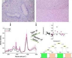
FT-Raman spectra in combination with machine learning and multivariate analyses as a diagnostic tool in brain tumors
Brain tumors are one of the most dangerous, because the position of these are in the organ that governs all life processes. Moreover, a lot of brain tumor types were observed, but only one main diagnostic method was used – histopathology, for which preparation of sample was long. Consequently, a new, quicker diagnostic method is needed. In this paper, FT-Raman spectra of brain tissues were analyzed by Principal Component Analysis (PCA), Hierarchical Cluster Analysis (HCA), four different machine learning (ML) algorithms to show possibility of differentiating between glioblastoma G4 and meningiomas, as well as two different types of meningiomas (atypical and angiomatous). Obtained results showed that in meningiomas additional peak around 1503 cm−1 and higher level of amides was noticed in comparison with glioblastoma G4. In the case of meningiomas differentiation, in angiomatous meningiomas tissues lower level of lipids and polysaccharides were visible than in atypical meningiomas. Moreover, PCA analyses showed higher distinction between glioblastoma G4 and meningiomas in the FT-Raman range between 800 cm−1 and 1800 cm−1 and between two types of meningiomas in the range between 2700 cm−1 and 3000 cm−1. Decision trees showed, that the most important peaks to differentiate glioblastoma and meningiomas were at 1151 cm−1 and 2836 cm−1 while for angiomatous and atypical meningiomas – 1514 cm−1 and 2875 cm−1. Furthermore, the accuracy of obtained results for glioblastoma G4 and meningiomas was 88 %, while for meningiomas – 92 %. Consequently, obtained data showed possibility of using FT-Raman spectroscopy in diagnosis of different types of brain tumors.
期刊介绍:
The mission of Nanomedicine: Nanotechnology, Biology, and Medicine (Nanomedicine: NBM) is to promote the emerging interdisciplinary field of nanomedicine.
Nanomedicine: NBM is an international, peer-reviewed journal presenting novel, significant, and interdisciplinary theoretical and experimental results related to nanoscience and nanotechnology in the life and health sciences. Content includes basic, translational, and clinical research addressing diagnosis, treatment, monitoring, prediction, and prevention of diseases.


 分享
分享
 求助内容:
求助内容: 应助结果提醒方式:
 扫码关注我们
