{"title":"利用基于体素的形态计量学和连接性分析法分析扩散峰度成像的白质结构差异。","authors":"Yuki Kanazawa, Natsuki Ikemitsu, Yuki Kinjo, Masafumi Harada, Hiroaki Hayashi, Yo Taniguchi, Kosuke Ito, Yoshitaka Bito, Yuki Matsumoto, Akihiro Haga","doi":"10.1093/bjro/tzad003","DOIUrl":null,"url":null,"abstract":"<p><strong>Objectives: </strong>In a clinical study, diffusion kurtosis imaging (DKI) has been used to visualize and distinguish white matter (WM) structures' details. The purpose of our study is to evaluate and compare the diffusion tensor imaging (DTI) and DKI parameter values to obtain WM structure differences of healthy subjects.</p><p><strong>Methods: </strong>Thirteen healthy volunteers (mean age, 25.2 years) were examined in this study. On a 3-T MRI system, diffusion dataset for DKI was acquired using an echo-planner imaging sequence, and T<sub>1</sub>-weghted (T<sub>1</sub>w) images were acquired. Imaging analysis was performed using Functional MRI of the brain Software Library (FSL). First, registration analysis was performed using the T<sub>1</sub>w of each subject to MNI152. Second, DTI (eg, fractional anisotropy [FA] and each diffusivity) and DKI (eg, mean kurtosis [MK], radial kurtosis [RK], and axial kurtosis [AK]) datasets were applied to above computed spline coefficients and affine matrices. Each DTI and DKI parameter value for WM areas was compared. Finally, tract-based spatial statistics (TBSS) analysis was performed using each parameter.</p><p><strong>Results: </strong>The relationship between FA and kurtosis parameters (MK, RK, and AK) for WM areas had a strong positive correlation (FA-MK, <i>R</i><sup>2</sup> = 0.93; FA-RK, <i>R</i><sup>2</sup> = 0.89) and a strong negative correlation (FA-AK, <i>R</i><sup>2</sup> = 0.92). When comparing a TBSS connection, we found that this could be observed more clearly in MK than in RK and FA.</p><p><strong>Conclusions: </strong>WM analysis with DKI enable us to obtain more detailed information for connectivity between nerve structures.</p><p><strong>Advances in knowledge: </strong>Quantitative indices of neurological diseases were determined using segmenting WM regions using voxel-based morphometry processing of DKI images.</p>","PeriodicalId":72419,"journal":{"name":"BJR open","volume":"6 1","pages":"tzad003"},"PeriodicalIF":2.1000,"publicationDate":"2023-12-12","publicationTypes":"Journal Article","fieldsOfStudy":null,"isOpenAccess":false,"openAccessPdf":"https://www.ncbi.nlm.nih.gov/pmc/articles/PMC10860519/pdf/","citationCount":"0","resultStr":"{\"title\":\"Differences of white matter structure for diffusion kurtosis imaging using voxel-based morphometry and connectivity analysis.\",\"authors\":\"Yuki Kanazawa, Natsuki Ikemitsu, Yuki Kinjo, Masafumi Harada, Hiroaki Hayashi, Yo Taniguchi, Kosuke Ito, Yoshitaka Bito, Yuki Matsumoto, Akihiro Haga\",\"doi\":\"10.1093/bjro/tzad003\",\"DOIUrl\":null,\"url\":null,\"abstract\":\"<p><strong>Objectives: </strong>In a clinical study, diffusion kurtosis imaging (DKI) has been used to visualize and distinguish white matter (WM) structures' details. The purpose of our study is to evaluate and compare the diffusion tensor imaging (DTI) and DKI parameter values to obtain WM structure differences of healthy subjects.</p><p><strong>Methods: </strong>Thirteen healthy volunteers (mean age, 25.2 years) were examined in this study. On a 3-T MRI system, diffusion dataset for DKI was acquired using an echo-planner imaging sequence, and T<sub>1</sub>-weghted (T<sub>1</sub>w) images were acquired. Imaging analysis was performed using Functional MRI of the brain Software Library (FSL). First, registration analysis was performed using the T<sub>1</sub>w of each subject to MNI152. Second, DTI (eg, fractional anisotropy [FA] and each diffusivity) and DKI (eg, mean kurtosis [MK], radial kurtosis [RK], and axial kurtosis [AK]) datasets were applied to above computed spline coefficients and affine matrices. Each DTI and DKI parameter value for WM areas was compared. Finally, tract-based spatial statistics (TBSS) analysis was performed using each parameter.</p><p><strong>Results: </strong>The relationship between FA and kurtosis parameters (MK, RK, and AK) for WM areas had a strong positive correlation (FA-MK, <i>R</i><sup>2</sup> = 0.93; FA-RK, <i>R</i><sup>2</sup> = 0.89) and a strong negative correlation (FA-AK, <i>R</i><sup>2</sup> = 0.92). When comparing a TBSS connection, we found that this could be observed more clearly in MK than in RK and FA.</p><p><strong>Conclusions: </strong>WM analysis with DKI enable us to obtain more detailed information for connectivity between nerve structures.</p><p><strong>Advances in knowledge: </strong>Quantitative indices of neurological diseases were determined using segmenting WM regions using voxel-based morphometry processing of DKI images.</p>\",\"PeriodicalId\":72419,\"journal\":{\"name\":\"BJR open\",\"volume\":\"6 1\",\"pages\":\"tzad003\"},\"PeriodicalIF\":2.1000,\"publicationDate\":\"2023-12-12\",\"publicationTypes\":\"Journal Article\",\"fieldsOfStudy\":null,\"isOpenAccess\":false,\"openAccessPdf\":\"https://www.ncbi.nlm.nih.gov/pmc/articles/PMC10860519/pdf/\",\"citationCount\":\"0\",\"resultStr\":null,\"platform\":\"Semanticscholar\",\"paperid\":null,\"PeriodicalName\":\"BJR open\",\"FirstCategoryId\":\"1085\",\"ListUrlMain\":\"https://doi.org/10.1093/bjro/tzad003\",\"RegionNum\":0,\"RegionCategory\":null,\"ArticlePicture\":[],\"TitleCN\":null,\"AbstractTextCN\":null,\"PMCID\":null,\"EPubDate\":\"2024/1/1 0:00:00\",\"PubModel\":\"eCollection\",\"JCR\":\"\",\"JCRName\":\"\",\"Score\":null,\"Total\":0}","platform":"Semanticscholar","paperid":null,"PeriodicalName":"BJR open","FirstCategoryId":"1085","ListUrlMain":"https://doi.org/10.1093/bjro/tzad003","RegionNum":0,"RegionCategory":null,"ArticlePicture":[],"TitleCN":null,"AbstractTextCN":null,"PMCID":null,"EPubDate":"2024/1/1 0:00:00","PubModel":"eCollection","JCR":"","JCRName":"","Score":null,"Total":0}
引用次数: 0
摘要
目的:在临床研究中,弥散峰度成像(DKI)被用于观察和区分白质(WM)结构的细节。我们的研究旨在评估和比较弥散张量成像(DTI)和 DKI 参数值,以获得健康受试者白质结构的差异:本研究对 13 名健康志愿者(平均年龄 25.2 岁)进行了检查。在 3-T 磁共振成像系统上,使用回声扫描仪成像序列获取 DKI 扩散数据集,并获取 T1 加权(T1w)图像。使用大脑功能磁共振成像软件库(FSL)进行成像分析。首先,使用每个受试者的 T1w 与 MNI152 进行配准分析。其次,将 DTI(如分数各向异性[FA]和各扩散率)和 DKI(如平均峰度[MK]、径向峰度[RK]和轴向峰度[AK])数据集应用于上述计算出的样条系数和仿射矩阵。对 WM 区域的每个 DTI 和 DKI 参数值进行了比较。最后,利用每个参数进行了基于束的空间统计(TBSS)分析:结果:WM 区域的 FA 和峰度参数(MK、RK 和 AK)之间的关系具有很强的正相关性(FA-MK,R2 = 0.93;FA-RK,R2 = 0.89)和很强的负相关性(FA-AK,R2 = 0.92)。在比较 TBSS 连接时,我们发现在 MK 中比在 RK 和 FA 中能更清楚地观察到这一点:结论:通过 DKI 进行 WM 分析,我们可以获得神经结构之间连接的更详细信息:通过对 DKI 图像进行基于体素的形态计量学处理,对 WM 区域进行分割,从而确定神经系统疾病的定量指标。
Differences of white matter structure for diffusion kurtosis imaging using voxel-based morphometry and connectivity analysis.
Objectives: In a clinical study, diffusion kurtosis imaging (DKI) has been used to visualize and distinguish white matter (WM) structures' details. The purpose of our study is to evaluate and compare the diffusion tensor imaging (DTI) and DKI parameter values to obtain WM structure differences of healthy subjects.
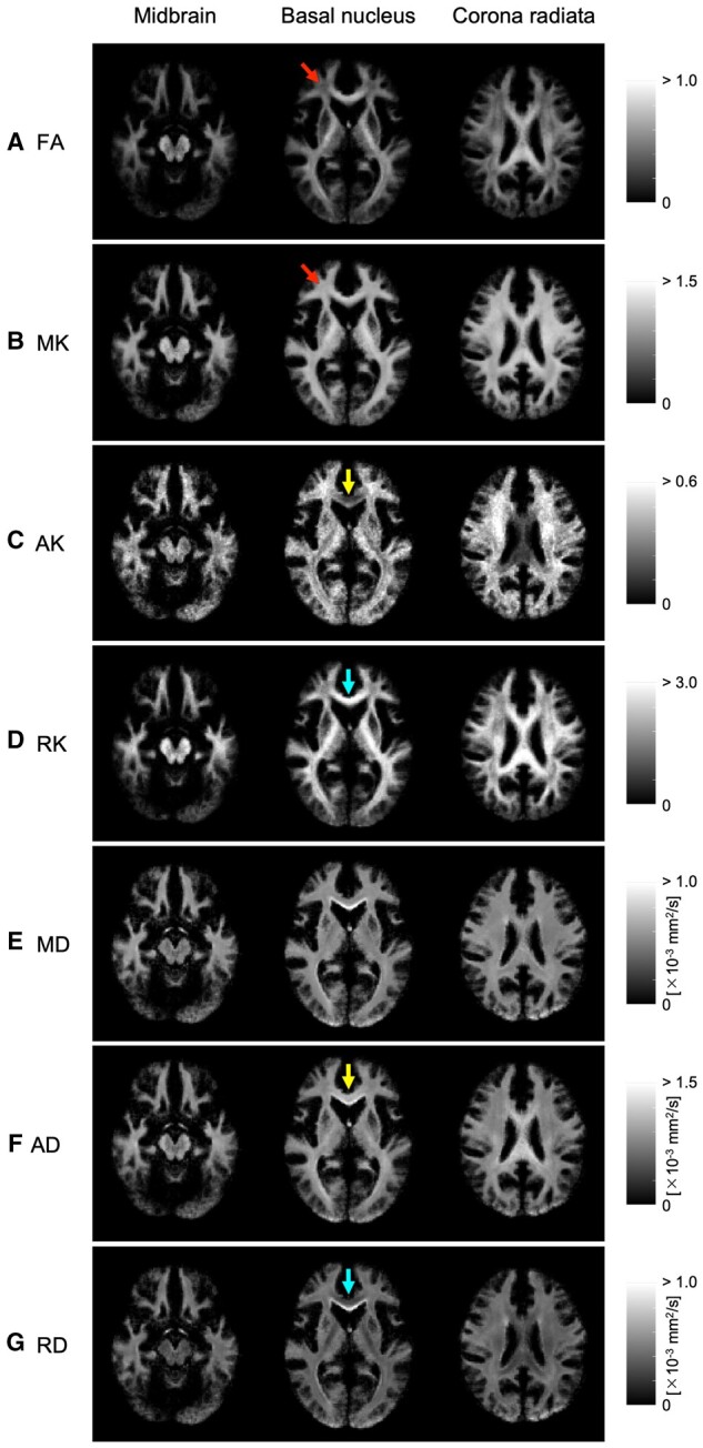
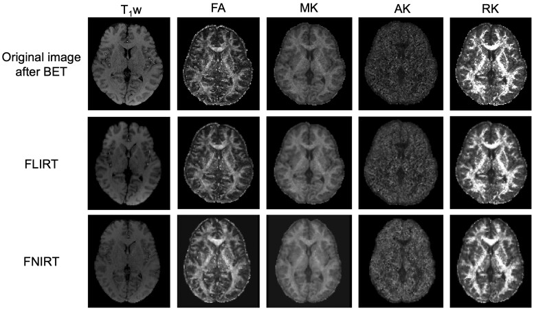
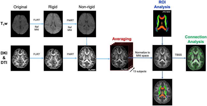
Methods: Thirteen healthy volunteers (mean age, 25.2 years) were examined in this study. On a 3-T MRI system, diffusion dataset for DKI was acquired using an echo-planner imaging sequence, and T1-weghted (T1w) images were acquired. Imaging analysis was performed using Functional MRI of the brain Software Library (FSL). First, registration analysis was performed using the T1w of each subject to MNI152. Second, DTI (eg, fractional anisotropy [FA] and each diffusivity) and DKI (eg, mean kurtosis [MK], radial kurtosis [RK], and axial kurtosis [AK]) datasets were applied to above computed spline coefficients and affine matrices. Each DTI and DKI parameter value for WM areas was compared. Finally, tract-based spatial statistics (TBSS) analysis was performed using each parameter.
Results: The relationship between FA and kurtosis parameters (MK, RK, and AK) for WM areas had a strong positive correlation (FA-MK, R2 = 0.93; FA-RK, R2 = 0.89) and a strong negative correlation (FA-AK, R2 = 0.92). When comparing a TBSS connection, we found that this could be observed more clearly in MK than in RK and FA.
Conclusions: WM analysis with DKI enable us to obtain more detailed information for connectivity between nerve structures.
Advances in knowledge: Quantitative indices of neurological diseases were determined using segmenting WM regions using voxel-based morphometry processing of DKI images.


 分享
分享

 求助内容:
求助内容: 应助结果提醒方式:
 扫码关注我们
