Marco Parillo, Carlo Augusto Mallio, Ilona A. Dekkers, Àlex Rovira, Aart J. van der Molen, Carlo Cosimo Quattrocchi
{"title":"静脉注射细胞外钆基造影剂后核磁共振成像中的钆增强延迟/延迟:值得等待吗?","authors":"Marco Parillo, Carlo Augusto Mallio, Ilona A. Dekkers, Àlex Rovira, Aart J. van der Molen, Carlo Cosimo Quattrocchi","doi":"10.1007/s10334-024-01151-0","DOIUrl":null,"url":null,"abstract":"<p>The acquisition of images minutes or even hours after intravenous extracellular gadolinium-based contrast agents (GBCA) administration (“Late/Delayed Gadolinium Enhancement” imaging; in this review, further termed LGE) has gained significant prominence in recent years in magnetic resonance imaging. The major limitation of LGE is the long examination time; thus, it becomes necessary to understand when it is worth waiting time after the intravenous injection of GBCA and which additional information comes from LGE. LGE can potentially be applied to various anatomical sites, such as heart, arterial vessels, lung, brain, abdomen, breast, and the musculoskeletal system, with different pathophysiological mechanisms. One of the most popular clinical applications of LGE regards the assessment of myocardial tissue thanks to its ability to highlight areas of acute myocardial damage and fibrotic tissues. Other frequently applied clinical contexts involve the study of the urinary tract with magnetic resonance urography and identifying pathological abdominal processes characterized by high fibrous stroma, such as biliary tract tumors, autoimmune pancreatitis, or intestinal fibrosis in Crohn’s disease. One of the current areas of heightened research interest revolves around the possibility of non-invasively studying the dynamics of neurofluids in the brain (the glymphatic system), the disruption of which could underlie many neurological disorders.</p>","PeriodicalId":18067,"journal":{"name":"Magnetic Resonance Materials in Physics, Biology and Medicine","volume":"68 1","pages":""},"PeriodicalIF":2.5000,"publicationDate":"2024-02-22","publicationTypes":"Journal Article","fieldsOfStudy":null,"isOpenAccess":false,"openAccessPdf":"","citationCount":"0","resultStr":"{\"title\":\"Late/delayed gadolinium enhancement in MRI after intravenous administration of extracellular gadolinium-based contrast agents: is it worth waiting?\",\"authors\":\"Marco Parillo, Carlo Augusto Mallio, Ilona A. Dekkers, Àlex Rovira, Aart J. van der Molen, Carlo Cosimo Quattrocchi\",\"doi\":\"10.1007/s10334-024-01151-0\",\"DOIUrl\":null,\"url\":null,\"abstract\":\"<p>The acquisition of images minutes or even hours after intravenous extracellular gadolinium-based contrast agents (GBCA) administration (“Late/Delayed Gadolinium Enhancement” imaging; in this review, further termed LGE) has gained significant prominence in recent years in magnetic resonance imaging. The major limitation of LGE is the long examination time; thus, it becomes necessary to understand when it is worth waiting time after the intravenous injection of GBCA and which additional information comes from LGE. LGE can potentially be applied to various anatomical sites, such as heart, arterial vessels, lung, brain, abdomen, breast, and the musculoskeletal system, with different pathophysiological mechanisms. One of the most popular clinical applications of LGE regards the assessment of myocardial tissue thanks to its ability to highlight areas of acute myocardial damage and fibrotic tissues. Other frequently applied clinical contexts involve the study of the urinary tract with magnetic resonance urography and identifying pathological abdominal processes characterized by high fibrous stroma, such as biliary tract tumors, autoimmune pancreatitis, or intestinal fibrosis in Crohn’s disease. One of the current areas of heightened research interest revolves around the possibility of non-invasively studying the dynamics of neurofluids in the brain (the glymphatic system), the disruption of which could underlie many neurological disorders.</p>\",\"PeriodicalId\":18067,\"journal\":{\"name\":\"Magnetic Resonance Materials in Physics, Biology and Medicine\",\"volume\":\"68 1\",\"pages\":\"\"},\"PeriodicalIF\":2.5000,\"publicationDate\":\"2024-02-22\",\"publicationTypes\":\"Journal Article\",\"fieldsOfStudy\":null,\"isOpenAccess\":false,\"openAccessPdf\":\"\",\"citationCount\":\"0\",\"resultStr\":null,\"platform\":\"Semanticscholar\",\"paperid\":null,\"PeriodicalName\":\"Magnetic Resonance Materials in Physics, Biology and Medicine\",\"FirstCategoryId\":\"3\",\"ListUrlMain\":\"https://doi.org/10.1007/s10334-024-01151-0\",\"RegionNum\":4,\"RegionCategory\":\"医学\",\"ArticlePicture\":[],\"TitleCN\":null,\"AbstractTextCN\":null,\"PMCID\":null,\"EPubDate\":\"\",\"PubModel\":\"\",\"JCR\":\"Q3\",\"JCRName\":\"RADIOLOGY, NUCLEAR MEDICINE & MEDICAL IMAGING\",\"Score\":null,\"Total\":0}","platform":"Semanticscholar","paperid":null,"PeriodicalName":"Magnetic Resonance Materials in Physics, Biology and Medicine","FirstCategoryId":"3","ListUrlMain":"https://doi.org/10.1007/s10334-024-01151-0","RegionNum":4,"RegionCategory":"医学","ArticlePicture":[],"TitleCN":null,"AbstractTextCN":null,"PMCID":null,"EPubDate":"","PubModel":"","JCR":"Q3","JCRName":"RADIOLOGY, NUCLEAR MEDICINE & MEDICAL IMAGING","Score":null,"Total":0}
Late/delayed gadolinium enhancement in MRI after intravenous administration of extracellular gadolinium-based contrast agents: is it worth waiting?
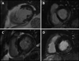
The acquisition of images minutes or even hours after intravenous extracellular gadolinium-based contrast agents (GBCA) administration (“Late/Delayed Gadolinium Enhancement” imaging; in this review, further termed LGE) has gained significant prominence in recent years in magnetic resonance imaging. The major limitation of LGE is the long examination time; thus, it becomes necessary to understand when it is worth waiting time after the intravenous injection of GBCA and which additional information comes from LGE. LGE can potentially be applied to various anatomical sites, such as heart, arterial vessels, lung, brain, abdomen, breast, and the musculoskeletal system, with different pathophysiological mechanisms. One of the most popular clinical applications of LGE regards the assessment of myocardial tissue thanks to its ability to highlight areas of acute myocardial damage and fibrotic tissues. Other frequently applied clinical contexts involve the study of the urinary tract with magnetic resonance urography and identifying pathological abdominal processes characterized by high fibrous stroma, such as biliary tract tumors, autoimmune pancreatitis, or intestinal fibrosis in Crohn’s disease. One of the current areas of heightened research interest revolves around the possibility of non-invasively studying the dynamics of neurofluids in the brain (the glymphatic system), the disruption of which could underlie many neurological disorders.
期刊介绍:
MAGMA is a multidisciplinary international journal devoted to the publication of articles on all aspects of magnetic resonance techniques and their applications in medicine and biology. MAGMA currently publishes research papers, reviews, letters to the editor, and commentaries, six times a year. The subject areas covered by MAGMA include:
advances in materials, hardware and software in magnetic resonance technology,
new developments and results in research and practical applications of magnetic resonance imaging and spectroscopy related to biology and medicine,
study of animal models and intact cells using magnetic resonance,
reports of clinical trials on humans and clinical validation of magnetic resonance protocols.


 分享
分享
 求助内容:
求助内容: 应助结果提醒方式:
 扫码关注我们
