Jeremy W. Mortimer, Philippa A. Rust, Jennifer Z. Paxton
{"title":"新型三维共培养系统的解剖学设计与制作(复制人体屈指深肌内侧)。","authors":"Jeremy W. Mortimer, Philippa A. Rust, Jennifer Z. Paxton","doi":"10.1111/joa.14027","DOIUrl":null,"url":null,"abstract":"<p>The enthesis, the specialized junction between tendon and bone, is a common site of injury. Although notoriously difficult to repair, advances in interfacial tissue engineering techniques are being developed for restorative function. Most notably are 3D in vitro co-culture models, built to recreate the complex heterogeneity of the native enthesis. While cell and matrix properties are often considered, there has been little attention given to native enthesis anatomical morphometrics and replicating these to enhance clinical relevance. This study focuses on the flexor digitorum profundus (FDP) tendon enthesis and, by combining anatomical morphometrics with computer-aided design, demonstrates the design and construction of an accurate and scalable model of the FDP enthesis. Bespoke 3D-printed mould inserts were fabricated based on the size, shape and insertion angle of the FDP enthesis. Then, silicone culture moulds were created, enabling the production of bespoke anatomical culture zones for an in vitro FDP enthesis model. The validity of the model has been confirmed using brushite cement scaffolds seeded with osteoblasts (bone) and fibrin hydrogel scaffolds seeded with fibroblasts (tendon) in individual studies with cells from either human or rat origin. This novel approach allows a bespoke anatomical design for enthesis repair and should be applied to future studies in this area.</p>","PeriodicalId":14971,"journal":{"name":"Journal of Anatomy","volume":"248 4","pages":"598-611"},"PeriodicalIF":1.9000,"publicationDate":"2026-03-19","publicationTypes":"Journal Article","fieldsOfStudy":null,"isOpenAccess":false,"openAccessPdf":"https://onlinelibrary.wiley.com/doi/epdf/10.1111/joa.14027","citationCount":"0","resultStr":"{\"title\":\"Anatomical design and production of a novel three-dimensional co-culture system replicating the human flexor digitorum profundus enthesis\",\"authors\":\"Jeremy W. Mortimer, Philippa A. Rust, Jennifer Z. Paxton\",\"doi\":\"10.1111/joa.14027\",\"DOIUrl\":null,\"url\":null,\"abstract\":\"<p>The enthesis, the specialized junction between tendon and bone, is a common site of injury. Although notoriously difficult to repair, advances in interfacial tissue engineering techniques are being developed for restorative function. Most notably are 3D in vitro co-culture models, built to recreate the complex heterogeneity of the native enthesis. While cell and matrix properties are often considered, there has been little attention given to native enthesis anatomical morphometrics and replicating these to enhance clinical relevance. This study focuses on the flexor digitorum profundus (FDP) tendon enthesis and, by combining anatomical morphometrics with computer-aided design, demonstrates the design and construction of an accurate and scalable model of the FDP enthesis. Bespoke 3D-printed mould inserts were fabricated based on the size, shape and insertion angle of the FDP enthesis. Then, silicone culture moulds were created, enabling the production of bespoke anatomical culture zones for an in vitro FDP enthesis model. The validity of the model has been confirmed using brushite cement scaffolds seeded with osteoblasts (bone) and fibrin hydrogel scaffolds seeded with fibroblasts (tendon) in individual studies with cells from either human or rat origin. This novel approach allows a bespoke anatomical design for enthesis repair and should be applied to future studies in this area.</p>\",\"PeriodicalId\":14971,\"journal\":{\"name\":\"Journal of Anatomy\",\"volume\":\"248 4\",\"pages\":\"598-611\"},\"PeriodicalIF\":1.9000,\"publicationDate\":\"2026-03-19\",\"publicationTypes\":\"Journal Article\",\"fieldsOfStudy\":null,\"isOpenAccess\":false,\"openAccessPdf\":\"https://onlinelibrary.wiley.com/doi/epdf/10.1111/joa.14027\",\"citationCount\":\"0\",\"resultStr\":null,\"platform\":\"Semanticscholar\",\"paperid\":null,\"PeriodicalName\":\"Journal of Anatomy\",\"FirstCategoryId\":\"3\",\"ListUrlMain\":\"https://onlinelibrary.wiley.com/doi/10.1111/joa.14027\",\"RegionNum\":3,\"RegionCategory\":\"医学\",\"ArticlePicture\":[],\"TitleCN\":null,\"AbstractTextCN\":null,\"PMCID\":null,\"EPubDate\":\"2024/2/23 0:00:00\",\"PubModel\":\"Epub\",\"JCR\":\"Q2\",\"JCRName\":\"ANATOMY & MORPHOLOGY\",\"Score\":null,\"Total\":0}","platform":"Semanticscholar","paperid":null,"PeriodicalName":"Journal of Anatomy","FirstCategoryId":"3","ListUrlMain":"https://onlinelibrary.wiley.com/doi/10.1111/joa.14027","RegionNum":3,"RegionCategory":"医学","ArticlePicture":[],"TitleCN":null,"AbstractTextCN":null,"PMCID":null,"EPubDate":"2024/2/23 0:00:00","PubModel":"Epub","JCR":"Q2","JCRName":"ANATOMY & MORPHOLOGY","Score":null,"Total":0}
引用次数: 0
摘要
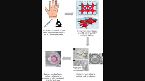
肌腱与骨骼之间的特殊交界处--肌腱内侧是常见的损伤部位。虽然修复难度很大,但界面组织工程技术的进步正在用于修复功能。最值得注意的是三维体外共培养模型,该模型的建立是为了再现原生内骨的复杂异质性。虽然细胞和基质的特性经常被考虑在内,但人们很少关注原生内植物的解剖形态和复制这些形态以提高临床相关性。本研究侧重于屈指深肌(FDP)肌腱内膜,通过将解剖形态计量学与计算机辅助设计相结合,展示了如何设计和构建精确且可扩展的 FDP 内膜模型。根据 FDP 肌腱假体的尺寸、形状和插入角度,制作了定制的 3D 打印模具插入件。然后,制作了硅胶培养模具,为体外 FDP 内植物模型量身定做了解剖培养区。在对人类或大鼠细胞进行的个别研究中,使用毛刷状水泥支架播种成骨细胞(骨)和纤维蛋白水凝胶支架播种成纤维细胞(肌腱),证实了该模型的有效性。这种新颖的方法可为假体修复提供定制的解剖学设计,应在该领域的未来研究中加以应用。
Anatomical design and production of a novel three-dimensional co-culture system replicating the human flexor digitorum profundus enthesis
The enthesis, the specialized junction between tendon and bone, is a common site of injury. Although notoriously difficult to repair, advances in interfacial tissue engineering techniques are being developed for restorative function. Most notably are 3D in vitro co-culture models, built to recreate the complex heterogeneity of the native enthesis. While cell and matrix properties are often considered, there has been little attention given to native enthesis anatomical morphometrics and replicating these to enhance clinical relevance. This study focuses on the flexor digitorum profundus (FDP) tendon enthesis and, by combining anatomical morphometrics with computer-aided design, demonstrates the design and construction of an accurate and scalable model of the FDP enthesis. Bespoke 3D-printed mould inserts were fabricated based on the size, shape and insertion angle of the FDP enthesis. Then, silicone culture moulds were created, enabling the production of bespoke anatomical culture zones for an in vitro FDP enthesis model. The validity of the model has been confirmed using brushite cement scaffolds seeded with osteoblasts (bone) and fibrin hydrogel scaffolds seeded with fibroblasts (tendon) in individual studies with cells from either human or rat origin. This novel approach allows a bespoke anatomical design for enthesis repair and should be applied to future studies in this area.
期刊介绍:
Journal of Anatomy is an international peer-reviewed journal sponsored by the Anatomical Society. The journal publishes original papers, invited review articles and book reviews. Its main focus is to understand anatomy through an analysis of structure, function, development and evolution. Priority will be given to studies of that clearly articulate their relevance to the anatomical community. Focal areas include: experimental studies, contributions based on molecular and cell biology and on the application of modern imaging techniques and papers with novel methods or synthetic perspective on an anatomical system.
Studies that are essentially descriptive anatomy are appropriate only if they communicate clearly a broader functional or evolutionary significance. You must clearly state the broader implications of your work in the abstract.
We particularly welcome submissions in the following areas:
Cell biology and tissue architecture
Comparative functional morphology
Developmental biology
Evolutionary developmental biology
Evolutionary morphology
Functional human anatomy
Integrative vertebrate paleontology
Methodological innovations in anatomical research
Musculoskeletal system
Neuroanatomy and neurodegeneration
Significant advances in anatomical education.


 分享
分享
 求助内容:
求助内容: 应助结果提醒方式:
 扫码关注我们
