Rhea L. Storlund, David A. S. Rosen, Martin Haulena, Shubhayan Sanatani, Jessica Vander Zaag, Andrew W. Trites
{"title":"麻醉后的北部海狗和斯特勒海狮的升主动脉超声波图像证实,主动脉球保持着持续的血流。","authors":"Rhea L. Storlund, David A. S. Rosen, Martin Haulena, Shubhayan Sanatani, Jessica Vander Zaag, Andrew W. Trites","doi":"10.1002/jez.2799","DOIUrl":null,"url":null,"abstract":"<p>The increased size and enhanced compliance of the aortic bulb—the enlargement of the ascending aorta—are believed to maintain blood flow in pinnipeds during extended periods of diastole induced by diving bradycardia. The aortic bulb has been described ex vivo in several species of pinnipeds, but in vivo measurements are needed to investigate the relationship between structure and function. We obtained ultrasound images using electrocardiogram-gated transesophageal echocardiography during anesthesia and after atropine administration to assess the relationship between aortic bulb anatomy and cardiac function (heart rate, stroke volume, cardiac output) in northern fur seals (<i>Callorhinus ursinus</i>) and Steller sea lions (<i>Eumetopias jubatus</i>). We observed that the aortic bulb in northern fur seals and Steller sea lions expands during systole and recoils over the entire diastolic period indicating that blood flow is maintained throughout the entire cardiac cycle as expected. The stroke volumes we measured in the fur seals and sea lions fit the values predicted based on body size in mammals and did not change with increased heart rates, suggesting that greater stroke volumes are not needed for aortic bulb function. Overall, our results suggest that peripheral vasoconstriction during diving is sufficient to modulate the volume of blood in the aortic bulb to ensure that flow lasts over the entire diastolic period. These results indicate that the shift of blood into the aortic bulb of pinnipeds is a fundamental mechanism caused by vasoconstriction while diving, highlighting the importance of this unique anatomical adaptation.</p>","PeriodicalId":15711,"journal":{"name":"Journal of experimental zoology. Part A, Ecological and integrative physiology","volume":"341 4","pages":"458-469"},"PeriodicalIF":1.4000,"publicationDate":"2024-02-26","publicationTypes":"Journal Article","fieldsOfStudy":null,"isOpenAccess":false,"openAccessPdf":"https://onlinelibrary.wiley.com/doi/epdf/10.1002/jez.2799","citationCount":"0","resultStr":"{\"title\":\"Ultrasound images of the ascending aorta of anesthetized northern fur seals and Steller sea lions confirm that the aortic bulb maintains continuous blood flow\",\"authors\":\"Rhea L. Storlund, David A. S. Rosen, Martin Haulena, Shubhayan Sanatani, Jessica Vander Zaag, Andrew W. Trites\",\"doi\":\"10.1002/jez.2799\",\"DOIUrl\":null,\"url\":null,\"abstract\":\"<p>The increased size and enhanced compliance of the aortic bulb—the enlargement of the ascending aorta—are believed to maintain blood flow in pinnipeds during extended periods of diastole induced by diving bradycardia. The aortic bulb has been described ex vivo in several species of pinnipeds, but in vivo measurements are needed to investigate the relationship between structure and function. We obtained ultrasound images using electrocardiogram-gated transesophageal echocardiography during anesthesia and after atropine administration to assess the relationship between aortic bulb anatomy and cardiac function (heart rate, stroke volume, cardiac output) in northern fur seals (<i>Callorhinus ursinus</i>) and Steller sea lions (<i>Eumetopias jubatus</i>). We observed that the aortic bulb in northern fur seals and Steller sea lions expands during systole and recoils over the entire diastolic period indicating that blood flow is maintained throughout the entire cardiac cycle as expected. The stroke volumes we measured in the fur seals and sea lions fit the values predicted based on body size in mammals and did not change with increased heart rates, suggesting that greater stroke volumes are not needed for aortic bulb function. Overall, our results suggest that peripheral vasoconstriction during diving is sufficient to modulate the volume of blood in the aortic bulb to ensure that flow lasts over the entire diastolic period. These results indicate that the shift of blood into the aortic bulb of pinnipeds is a fundamental mechanism caused by vasoconstriction while diving, highlighting the importance of this unique anatomical adaptation.</p>\",\"PeriodicalId\":15711,\"journal\":{\"name\":\"Journal of experimental zoology. Part A, Ecological and integrative physiology\",\"volume\":\"341 4\",\"pages\":\"458-469\"},\"PeriodicalIF\":1.4000,\"publicationDate\":\"2024-02-26\",\"publicationTypes\":\"Journal Article\",\"fieldsOfStudy\":null,\"isOpenAccess\":false,\"openAccessPdf\":\"https://onlinelibrary.wiley.com/doi/epdf/10.1002/jez.2799\",\"citationCount\":\"0\",\"resultStr\":null,\"platform\":\"Semanticscholar\",\"paperid\":null,\"PeriodicalName\":\"Journal of experimental zoology. Part A, Ecological and integrative physiology\",\"FirstCategoryId\":\"99\",\"ListUrlMain\":\"https://onlinelibrary.wiley.com/doi/10.1002/jez.2799\",\"RegionNum\":3,\"RegionCategory\":\"生物学\",\"ArticlePicture\":[],\"TitleCN\":null,\"AbstractTextCN\":null,\"PMCID\":null,\"EPubDate\":\"\",\"PubModel\":\"\",\"JCR\":\"Q1\",\"JCRName\":\"ZOOLOGY\",\"Score\":null,\"Total\":0}","platform":"Semanticscholar","paperid":null,"PeriodicalName":"Journal of experimental zoology. Part A, Ecological and integrative physiology","FirstCategoryId":"99","ListUrlMain":"https://onlinelibrary.wiley.com/doi/10.1002/jez.2799","RegionNum":3,"RegionCategory":"生物学","ArticlePicture":[],"TitleCN":null,"AbstractTextCN":null,"PMCID":null,"EPubDate":"","PubModel":"","JCR":"Q1","JCRName":"ZOOLOGY","Score":null,"Total":0}
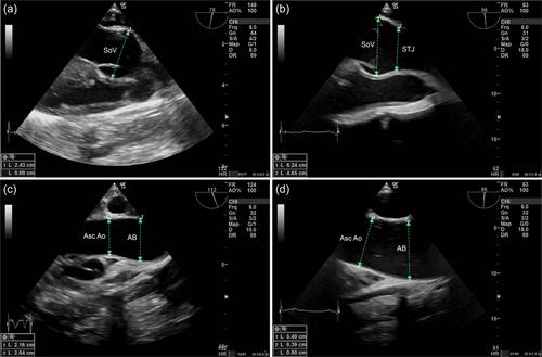
Ultrasound images of the ascending aorta of anesthetized northern fur seals and Steller sea lions confirm that the aortic bulb maintains continuous blood flow
The increased size and enhanced compliance of the aortic bulb—the enlargement of the ascending aorta—are believed to maintain blood flow in pinnipeds during extended periods of diastole induced by diving bradycardia. The aortic bulb has been described ex vivo in several species of pinnipeds, but in vivo measurements are needed to investigate the relationship between structure and function. We obtained ultrasound images using electrocardiogram-gated transesophageal echocardiography during anesthesia and after atropine administration to assess the relationship between aortic bulb anatomy and cardiac function (heart rate, stroke volume, cardiac output) in northern fur seals (Callorhinus ursinus) and Steller sea lions (Eumetopias jubatus). We observed that the aortic bulb in northern fur seals and Steller sea lions expands during systole and recoils over the entire diastolic period indicating that blood flow is maintained throughout the entire cardiac cycle as expected. The stroke volumes we measured in the fur seals and sea lions fit the values predicted based on body size in mammals and did not change with increased heart rates, suggesting that greater stroke volumes are not needed for aortic bulb function. Overall, our results suggest that peripheral vasoconstriction during diving is sufficient to modulate the volume of blood in the aortic bulb to ensure that flow lasts over the entire diastolic period. These results indicate that the shift of blood into the aortic bulb of pinnipeds is a fundamental mechanism caused by vasoconstriction while diving, highlighting the importance of this unique anatomical adaptation.
期刊介绍:
The Journal of Experimental Zoology – A publishes articles at the interface between Development, Physiology, Ecology and Evolution. Contributions that help to reveal how molecular, functional and ecological variation relate to one another are particularly welcome. The Journal publishes original research in the form of rapid communications or regular research articles, as well as perspectives and reviews on topics pertaining to the scope of the Journal. Acceptable articles are limited to studies on animals.


 分享
分享
 求助内容:
求助内容: 应助结果提醒方式:
 扫码关注我们
