Victor M Silva-Ortiz, Kenneth B Chapman, George C Chang Chien, Sudhir Diwan, Alaa Abd-Elsayed
{"title":"骶髂关节神经支配--针对骶骨侧支的新方法:实用方法。","authors":"Victor M Silva-Ortiz, Kenneth B Chapman, George C Chang Chien, Sudhir Diwan, Alaa Abd-Elsayed","doi":"10.1007/s40122-024-00585-7","DOIUrl":null,"url":null,"abstract":"<p><strong>Introduction: </strong>Sacroiliac joint (SIJ) pain is a relatively common cause of low back pain. Percutaneous radiofrequency (RF) techniques for SIJ are limited to ablation of the posterior SIJ innervation. Different techniques have been described for SIJ radiofrequency ablation, including conventional thermal, cooled RF, pulsed RF, bipolar RF, and specialized tip RF needle (i.e., multi-tined); however, additional costs may limit these applications.</p><p><strong>Methods: </strong>This new technique for SIJ denervation uses anatomical landmarks and a single RF cannula. Two spinal needles are placed lateral to the posterior S1 and S2 sacral foramina; then, with caudal tilt we get a coaxial view of the sacral bone, we advance an 18-G curved 15-mm active tip RF cannula just lateral to the aligned finder needles. Ablation is performed, and then the RF cannula is retracted 2 cm and ablation is repeated for a total of four lesions.</p><p><strong>Results: </strong>The two spinal needles placed lateral to the posterior sacral foramina S1 and S2 guide the final needle in the posterior aspect of the sacrum, lateral to the sacral foramina, where the lateral sacral branches are located.</p><p><strong>Conclusion: </strong>We introduce a cost and time efficient technique to perform radiofrequency ablation of the sacral lateral branches using a single RF needle. This technique utilizes the sacrum's reliable anatomy and angulation and maximizes the surface area of the active tip lesioning. This technique creates a strip lesion lateral to the sacral foramina and reduces time and cost efficacy compared to several of the other techniques and/or commercially available special devices designed for sacroiliac denervation.</p>","PeriodicalId":19908,"journal":{"name":"Pain and Therapy","volume":" ","pages":"281-286"},"PeriodicalIF":3.3000,"publicationDate":"2024-04-01","publicationTypes":"Journal Article","fieldsOfStudy":null,"isOpenAccess":false,"openAccessPdf":"https://www.ncbi.nlm.nih.gov/pmc/articles/PMC10928047/pdf/","citationCount":"0","resultStr":"{\"title\":\"Sacroiliac Joint Denervation-A Novel Approach to Target Sacral Lateral Branches: A Practical Approach.\",\"authors\":\"Victor M Silva-Ortiz, Kenneth B Chapman, George C Chang Chien, Sudhir Diwan, Alaa Abd-Elsayed\",\"doi\":\"10.1007/s40122-024-00585-7\",\"DOIUrl\":null,\"url\":null,\"abstract\":\"<p><strong>Introduction: </strong>Sacroiliac joint (SIJ) pain is a relatively common cause of low back pain. Percutaneous radiofrequency (RF) techniques for SIJ are limited to ablation of the posterior SIJ innervation. Different techniques have been described for SIJ radiofrequency ablation, including conventional thermal, cooled RF, pulsed RF, bipolar RF, and specialized tip RF needle (i.e., multi-tined); however, additional costs may limit these applications.</p><p><strong>Methods: </strong>This new technique for SIJ denervation uses anatomical landmarks and a single RF cannula. Two spinal needles are placed lateral to the posterior S1 and S2 sacral foramina; then, with caudal tilt we get a coaxial view of the sacral bone, we advance an 18-G curved 15-mm active tip RF cannula just lateral to the aligned finder needles. Ablation is performed, and then the RF cannula is retracted 2 cm and ablation is repeated for a total of four lesions.</p><p><strong>Results: </strong>The two spinal needles placed lateral to the posterior sacral foramina S1 and S2 guide the final needle in the posterior aspect of the sacrum, lateral to the sacral foramina, where the lateral sacral branches are located.</p><p><strong>Conclusion: </strong>We introduce a cost and time efficient technique to perform radiofrequency ablation of the sacral lateral branches using a single RF needle. This technique utilizes the sacrum's reliable anatomy and angulation and maximizes the surface area of the active tip lesioning. This technique creates a strip lesion lateral to the sacral foramina and reduces time and cost efficacy compared to several of the other techniques and/or commercially available special devices designed for sacroiliac denervation.</p>\",\"PeriodicalId\":19908,\"journal\":{\"name\":\"Pain and Therapy\",\"volume\":\" \",\"pages\":\"281-286\"},\"PeriodicalIF\":3.3000,\"publicationDate\":\"2024-04-01\",\"publicationTypes\":\"Journal Article\",\"fieldsOfStudy\":null,\"isOpenAccess\":false,\"openAccessPdf\":\"https://www.ncbi.nlm.nih.gov/pmc/articles/PMC10928047/pdf/\",\"citationCount\":\"0\",\"resultStr\":null,\"platform\":\"Semanticscholar\",\"paperid\":null,\"PeriodicalName\":\"Pain and Therapy\",\"FirstCategoryId\":\"3\",\"ListUrlMain\":\"https://doi.org/10.1007/s40122-024-00585-7\",\"RegionNum\":2,\"RegionCategory\":\"医学\",\"ArticlePicture\":[],\"TitleCN\":null,\"AbstractTextCN\":null,\"PMCID\":null,\"EPubDate\":\"2024/2/24 0:00:00\",\"PubModel\":\"Epub\",\"JCR\":\"Q1\",\"JCRName\":\"CLINICAL NEUROLOGY\",\"Score\":null,\"Total\":0}","platform":"Semanticscholar","paperid":null,"PeriodicalName":"Pain and Therapy","FirstCategoryId":"3","ListUrlMain":"https://doi.org/10.1007/s40122-024-00585-7","RegionNum":2,"RegionCategory":"医学","ArticlePicture":[],"TitleCN":null,"AbstractTextCN":null,"PMCID":null,"EPubDate":"2024/2/24 0:00:00","PubModel":"Epub","JCR":"Q1","JCRName":"CLINICAL NEUROLOGY","Score":null,"Total":0}
Sacroiliac Joint Denervation-A Novel Approach to Target Sacral Lateral Branches: A Practical Approach.
Introduction: Sacroiliac joint (SIJ) pain is a relatively common cause of low back pain. Percutaneous radiofrequency (RF) techniques for SIJ are limited to ablation of the posterior SIJ innervation. Different techniques have been described for SIJ radiofrequency ablation, including conventional thermal, cooled RF, pulsed RF, bipolar RF, and specialized tip RF needle (i.e., multi-tined); however, additional costs may limit these applications.
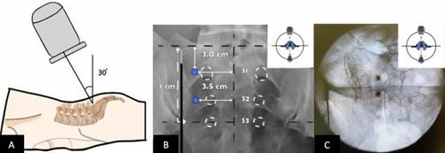
Methods: This new technique for SIJ denervation uses anatomical landmarks and a single RF cannula. Two spinal needles are placed lateral to the posterior S1 and S2 sacral foramina; then, with caudal tilt we get a coaxial view of the sacral bone, we advance an 18-G curved 15-mm active tip RF cannula just lateral to the aligned finder needles. Ablation is performed, and then the RF cannula is retracted 2 cm and ablation is repeated for a total of four lesions.
Results: The two spinal needles placed lateral to the posterior sacral foramina S1 and S2 guide the final needle in the posterior aspect of the sacrum, lateral to the sacral foramina, where the lateral sacral branches are located.
Conclusion: We introduce a cost and time efficient technique to perform radiofrequency ablation of the sacral lateral branches using a single RF needle. This technique utilizes the sacrum's reliable anatomy and angulation and maximizes the surface area of the active tip lesioning. This technique creates a strip lesion lateral to the sacral foramina and reduces time and cost efficacy compared to several of the other techniques and/or commercially available special devices designed for sacroiliac denervation.
期刊介绍:
Pain and Therapy is an international, open access, peer-reviewed, rapid publication journal dedicated to the publication of high-quality clinical (all phases), observational, real-world, and health outcomes research around the discovery, development, and use of pain therapies and pain-related devices. Studies relating to diagnosis, pharmacoeconomics, public health, quality of life, and patient care, management, and education are also encouraged.
Areas of focus include, but are not limited to, acute pain, cancer pain, chronic pain, headache and migraine, neuropathic pain, opioids, palliative care and pain ethics, peri- and post-operative pain as well as rheumatic pain and fibromyalgia.
The journal is of interest to a broad audience of pharmaceutical and healthcare professionals and publishes original research, reviews, case reports, trial protocols, short communications such as commentaries and editorials, and letters. The journal is read by a global audience and receives submissions from around the world. Pain and Therapy will consider all scientifically sound research be it positive, confirmatory or negative data. Submissions are welcomed whether they relate to an international and/or a country-specific audience, something that is crucially important when researchers are trying to target more specific patient populations. This inclusive approach allows the journal to assist in the dissemination of all scientifically and ethically sound research.


 分享
分享
 求助内容:
求助内容: 应助结果提醒方式:
 扫码关注我们
