Sena Aksel, Amber Derpinghaus, Mei Cao, Yi Li, Gerald Cunha, Laurence Baskin
{"title":"发育中的胎儿阴茎和阴蒂的神经血管解剖。","authors":"Sena Aksel, Amber Derpinghaus, Mei Cao, Yi Li, Gerald Cunha, Laurence Baskin","doi":"10.1111/joa.14029","DOIUrl":null,"url":null,"abstract":"<p>The human penile and clitoral development begins from a morphologically indifferent genital tubercle. Under the influence of androgen, the genital tubercle forms the penis by forming a tubular urethra within the penile shaft. Without the effect of the androgen, the genital tubercle differentiates into the clitoris, and a lack of formation of the urethra within the clitoris is observed. Even though there are similarities during the development of the glans penis and glans clitoris, the complex canalization occurring along the penile shaft eventually leads to a morphological difference between the penis and clitoris. Based on the morphological differences, the main goal of this study was to define the vascular and neuronal anatomy of the developing penis and clitoris between 8 and 12 weeks of gestation using laser scanning confocal microscopy. Our results demonstrated there is a co-expression of CD31, which is an endothelial cell marker, and PGP9.5, which is a neuronal marker in the penis where the fusion is actively occurring at the ventral shaft. We also identified a unique anatomical structure for the first time, the clitoral ridge, which is a fetal structure running along the clitoral shaft in the vestibular groove. Contrary to previous anatomical findings which indicate that the neurovascular distribution in the developing penis and clitoris is similar, in this study, laser scanning confocal microscopy enabled us to demonstrate finer differences in the neurovascular anatomy between the penis and clitoris.</p>","PeriodicalId":14971,"journal":{"name":"Journal of Anatomy","volume":"245 1","pages":"35-49"},"PeriodicalIF":2.2000,"publicationDate":"2024-02-28","publicationTypes":"Journal Article","fieldsOfStudy":null,"isOpenAccess":false,"openAccessPdf":"https://onlinelibrary.wiley.com/doi/epdf/10.1111/joa.14029","citationCount":"0","resultStr":"{\"title\":\"Neurovascular anatomy of the developing human fetal penis and clitoris\",\"authors\":\"Sena Aksel, Amber Derpinghaus, Mei Cao, Yi Li, Gerald Cunha, Laurence Baskin\",\"doi\":\"10.1111/joa.14029\",\"DOIUrl\":null,\"url\":null,\"abstract\":\"<p>The human penile and clitoral development begins from a morphologically indifferent genital tubercle. Under the influence of androgen, the genital tubercle forms the penis by forming a tubular urethra within the penile shaft. Without the effect of the androgen, the genital tubercle differentiates into the clitoris, and a lack of formation of the urethra within the clitoris is observed. Even though there are similarities during the development of the glans penis and glans clitoris, the complex canalization occurring along the penile shaft eventually leads to a morphological difference between the penis and clitoris. Based on the morphological differences, the main goal of this study was to define the vascular and neuronal anatomy of the developing penis and clitoris between 8 and 12 weeks of gestation using laser scanning confocal microscopy. Our results demonstrated there is a co-expression of CD31, which is an endothelial cell marker, and PGP9.5, which is a neuronal marker in the penis where the fusion is actively occurring at the ventral shaft. We also identified a unique anatomical structure for the first time, the clitoral ridge, which is a fetal structure running along the clitoral shaft in the vestibular groove. Contrary to previous anatomical findings which indicate that the neurovascular distribution in the developing penis and clitoris is similar, in this study, laser scanning confocal microscopy enabled us to demonstrate finer differences in the neurovascular anatomy between the penis and clitoris.</p>\",\"PeriodicalId\":14971,\"journal\":{\"name\":\"Journal of Anatomy\",\"volume\":\"245 1\",\"pages\":\"35-49\"},\"PeriodicalIF\":2.2000,\"publicationDate\":\"2024-02-28\",\"publicationTypes\":\"Journal Article\",\"fieldsOfStudy\":null,\"isOpenAccess\":false,\"openAccessPdf\":\"https://onlinelibrary.wiley.com/doi/epdf/10.1111/joa.14029\",\"citationCount\":\"0\",\"resultStr\":null,\"platform\":\"Semanticscholar\",\"paperid\":null,\"PeriodicalName\":\"Journal of Anatomy\",\"FirstCategoryId\":\"3\",\"ListUrlMain\":\"https://onlinelibrary.wiley.com/doi/10.1111/joa.14029\",\"RegionNum\":3,\"RegionCategory\":\"医学\",\"ArticlePicture\":[],\"TitleCN\":null,\"AbstractTextCN\":null,\"PMCID\":null,\"EPubDate\":\"\",\"PubModel\":\"\",\"JCR\":\"Q2\",\"JCRName\":\"ANATOMY & MORPHOLOGY\",\"Score\":null,\"Total\":0}","platform":"Semanticscholar","paperid":null,"PeriodicalName":"Journal of Anatomy","FirstCategoryId":"3","ListUrlMain":"https://onlinelibrary.wiley.com/doi/10.1111/joa.14029","RegionNum":3,"RegionCategory":"医学","ArticlePicture":[],"TitleCN":null,"AbstractTextCN":null,"PMCID":null,"EPubDate":"","PubModel":"","JCR":"Q2","JCRName":"ANATOMY & MORPHOLOGY","Score":null,"Total":0}
Neurovascular anatomy of the developing human fetal penis and clitoris
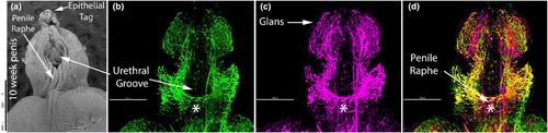
The human penile and clitoral development begins from a morphologically indifferent genital tubercle. Under the influence of androgen, the genital tubercle forms the penis by forming a tubular urethra within the penile shaft. Without the effect of the androgen, the genital tubercle differentiates into the clitoris, and a lack of formation of the urethra within the clitoris is observed. Even though there are similarities during the development of the glans penis and glans clitoris, the complex canalization occurring along the penile shaft eventually leads to a morphological difference between the penis and clitoris. Based on the morphological differences, the main goal of this study was to define the vascular and neuronal anatomy of the developing penis and clitoris between 8 and 12 weeks of gestation using laser scanning confocal microscopy. Our results demonstrated there is a co-expression of CD31, which is an endothelial cell marker, and PGP9.5, which is a neuronal marker in the penis where the fusion is actively occurring at the ventral shaft. We also identified a unique anatomical structure for the first time, the clitoral ridge, which is a fetal structure running along the clitoral shaft in the vestibular groove. Contrary to previous anatomical findings which indicate that the neurovascular distribution in the developing penis and clitoris is similar, in this study, laser scanning confocal microscopy enabled us to demonstrate finer differences in the neurovascular anatomy between the penis and clitoris.
期刊介绍:
Journal of Anatomy is an international peer-reviewed journal sponsored by the Anatomical Society. The journal publishes original papers, invited review articles and book reviews. Its main focus is to understand anatomy through an analysis of structure, function, development and evolution. Priority will be given to studies of that clearly articulate their relevance to the anatomical community. Focal areas include: experimental studies, contributions based on molecular and cell biology and on the application of modern imaging techniques and papers with novel methods or synthetic perspective on an anatomical system.
Studies that are essentially descriptive anatomy are appropriate only if they communicate clearly a broader functional or evolutionary significance. You must clearly state the broader implications of your work in the abstract.
We particularly welcome submissions in the following areas:
Cell biology and tissue architecture
Comparative functional morphology
Developmental biology
Evolutionary developmental biology
Evolutionary morphology
Functional human anatomy
Integrative vertebrate paleontology
Methodological innovations in anatomical research
Musculoskeletal system
Neuroanatomy and neurodegeneration
Significant advances in anatomical education.


 分享
分享
 求助内容:
求助内容: 应助结果提醒方式:
 扫码关注我们
