Michael H. Alexander, William J. Cousins, Tom Ewen, Andrew P. South, Penny Lovat, Niki Stefanos
{"title":"AMBRA1 和 SQSTM1 的联合免疫组化表达可识别有转移风险的分化不良皮肤鳞状细胞癌患者:概念验证研究。","authors":"Michael H. Alexander, William J. Cousins, Tom Ewen, Andrew P. South, Penny Lovat, Niki Stefanos","doi":"10.1111/cup.14590","DOIUrl":null,"url":null,"abstract":"<div>\n \n \n <section>\n \n <h3> Background</h3>\n \n <p>Cutaneous squamous cell carcinoma (cSCC) incidence continues to increase globally with, as of yet, an unmet need for reliable prognostic biomarkers to identify patients at increased risk of metastasis. The aim of the present study was to test the prognostic potential of the combined immunohistochemical expression of the autophagy regulatory biomarkers, AMBRA1 and SQSTM1, to identify high-risk patient subsets.</p>\n </section>\n \n <section>\n \n <h3> Methods</h3>\n \n <p>A retrospective cohort of 68 formalin-fixed paraffin-embedded primary cSCCs with known 5-year metastatic outcomes were subjected to automated immunohistochemical staining for AMBRA1 and SQSTM1. Digital images of stained slides were annotated to define four regions of interest: the normal and peritumoral epidermis, the tumor mass, and the tumor growth front. H-score analysis was used to semi-quantify AMBRA1 or SQSTM1 expression in each region of interest using Aperio ImageScope software, with receiver operator characteristics and Kaplan–Meier analysis used to assess prognostic potential.</p>\n </section>\n \n <section>\n \n <h3> Results</h3>\n \n <p>The combined loss of expression of AMBRA1 in the tumor growth front and SQSTM1 in the peritumoral epidermis identified patients with poorly differentiated cSCCs at risk of metastasis (*<i>p</i> < 0.05).</p>\n </section>\n \n <section>\n \n <h3> Conclusions</h3>\n \n <p>Collectively, these proof of concept data suggest loss of the combined expression of AMBRA1 in the cSCC growth front and SQSTM1 in the peritumoral epidermis as a putative prognostic biomarker for poorly differentiated cSCC.</p>\n </section>\n </div>","PeriodicalId":15407,"journal":{"name":"Journal of Cutaneous Pathology","volume":"51 6","pages":"450-458"},"PeriodicalIF":1.1000,"publicationDate":"2024-02-29","publicationTypes":"Journal Article","fieldsOfStudy":null,"isOpenAccess":false,"openAccessPdf":"https://onlinelibrary.wiley.com/doi/epdf/10.1111/cup.14590","citationCount":"0","resultStr":"{\"title\":\"The combined immunohistochemical expression of AMBRA1 and SQSTM1 identifies patients with poorly differentiated cutaneous squamous cell carcinoma at risk of metastasis: A proof of concept study\",\"authors\":\"Michael H. Alexander, William J. Cousins, Tom Ewen, Andrew P. South, Penny Lovat, Niki Stefanos\",\"doi\":\"10.1111/cup.14590\",\"DOIUrl\":null,\"url\":null,\"abstract\":\"<div>\\n \\n \\n <section>\\n \\n <h3> Background</h3>\\n \\n <p>Cutaneous squamous cell carcinoma (cSCC) incidence continues to increase globally with, as of yet, an unmet need for reliable prognostic biomarkers to identify patients at increased risk of metastasis. The aim of the present study was to test the prognostic potential of the combined immunohistochemical expression of the autophagy regulatory biomarkers, AMBRA1 and SQSTM1, to identify high-risk patient subsets.</p>\\n </section>\\n \\n <section>\\n \\n <h3> Methods</h3>\\n \\n <p>A retrospective cohort of 68 formalin-fixed paraffin-embedded primary cSCCs with known 5-year metastatic outcomes were subjected to automated immunohistochemical staining for AMBRA1 and SQSTM1. Digital images of stained slides were annotated to define four regions of interest: the normal and peritumoral epidermis, the tumor mass, and the tumor growth front. H-score analysis was used to semi-quantify AMBRA1 or SQSTM1 expression in each region of interest using Aperio ImageScope software, with receiver operator characteristics and Kaplan–Meier analysis used to assess prognostic potential.</p>\\n </section>\\n \\n <section>\\n \\n <h3> Results</h3>\\n \\n <p>The combined loss of expression of AMBRA1 in the tumor growth front and SQSTM1 in the peritumoral epidermis identified patients with poorly differentiated cSCCs at risk of metastasis (*<i>p</i> < 0.05).</p>\\n </section>\\n \\n <section>\\n \\n <h3> Conclusions</h3>\\n \\n <p>Collectively, these proof of concept data suggest loss of the combined expression of AMBRA1 in the cSCC growth front and SQSTM1 in the peritumoral epidermis as a putative prognostic biomarker for poorly differentiated cSCC.</p>\\n </section>\\n </div>\",\"PeriodicalId\":15407,\"journal\":{\"name\":\"Journal of Cutaneous Pathology\",\"volume\":\"51 6\",\"pages\":\"450-458\"},\"PeriodicalIF\":1.1000,\"publicationDate\":\"2024-02-29\",\"publicationTypes\":\"Journal Article\",\"fieldsOfStudy\":null,\"isOpenAccess\":false,\"openAccessPdf\":\"https://onlinelibrary.wiley.com/doi/epdf/10.1111/cup.14590\",\"citationCount\":\"0\",\"resultStr\":null,\"platform\":\"Semanticscholar\",\"paperid\":null,\"PeriodicalName\":\"Journal of Cutaneous Pathology\",\"FirstCategoryId\":\"3\",\"ListUrlMain\":\"https://onlinelibrary.wiley.com/doi/10.1111/cup.14590\",\"RegionNum\":4,\"RegionCategory\":\"医学\",\"ArticlePicture\":[],\"TitleCN\":null,\"AbstractTextCN\":null,\"PMCID\":null,\"EPubDate\":\"\",\"PubModel\":\"\",\"JCR\":\"Q3\",\"JCRName\":\"DERMATOLOGY\",\"Score\":null,\"Total\":0}","platform":"Semanticscholar","paperid":null,"PeriodicalName":"Journal of Cutaneous Pathology","FirstCategoryId":"3","ListUrlMain":"https://onlinelibrary.wiley.com/doi/10.1111/cup.14590","RegionNum":4,"RegionCategory":"医学","ArticlePicture":[],"TitleCN":null,"AbstractTextCN":null,"PMCID":null,"EPubDate":"","PubModel":"","JCR":"Q3","JCRName":"DERMATOLOGY","Score":null,"Total":0}
The combined immunohistochemical expression of AMBRA1 and SQSTM1 identifies patients with poorly differentiated cutaneous squamous cell carcinoma at risk of metastasis: A proof of concept study
Background
Cutaneous squamous cell carcinoma (cSCC) incidence continues to increase globally with, as of yet, an unmet need for reliable prognostic biomarkers to identify patients at increased risk of metastasis. The aim of the present study was to test the prognostic potential of the combined immunohistochemical expression of the autophagy regulatory biomarkers, AMBRA1 and SQSTM1, to identify high-risk patient subsets.
Methods
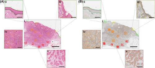
A retrospective cohort of 68 formalin-fixed paraffin-embedded primary cSCCs with known 5-year metastatic outcomes were subjected to automated immunohistochemical staining for AMBRA1 and SQSTM1. Digital images of stained slides were annotated to define four regions of interest: the normal and peritumoral epidermis, the tumor mass, and the tumor growth front. H-score analysis was used to semi-quantify AMBRA1 or SQSTM1 expression in each region of interest using Aperio ImageScope software, with receiver operator characteristics and Kaplan–Meier analysis used to assess prognostic potential.
Results
The combined loss of expression of AMBRA1 in the tumor growth front and SQSTM1 in the peritumoral epidermis identified patients with poorly differentiated cSCCs at risk of metastasis (*p < 0.05).
Conclusions
Collectively, these proof of concept data suggest loss of the combined expression of AMBRA1 in the cSCC growth front and SQSTM1 in the peritumoral epidermis as a putative prognostic biomarker for poorly differentiated cSCC.
期刊介绍:
Journal of Cutaneous Pathology publishes manuscripts broadly relevant to diseases of the skin and mucosae, with the aims of advancing scientific knowledge regarding dermatopathology and enhancing the communication between clinical practitioners and research scientists. Original scientific manuscripts on diagnostic and experimental cutaneous pathology are especially desirable. Timely, pertinent review articles also will be given high priority. Manuscripts based on light, fluorescence, and electron microscopy, histochemistry, immunology, molecular biology, and genetics, as well as allied sciences, are all welcome, provided their principal focus is on cutaneous pathology. Publication time will be kept as short as possible, ensuring that articles will be quickly available to all interested in this speciality.


 分享
分享
 求助内容:
求助内容: 应助结果提醒方式:
 扫码关注我们
