Tiankai Di, Chao Feng, Lulu Wang, Jinlong Xu, Yang Du, Baixiang Cheng, Yujiang Chen, Lian Wu
{"title":"增强牙髓发育过程中的血管生成:通过 FN1-ITGA5 信号传递的 DPSCs-ECs 通信","authors":"Tiankai Di, Chao Feng, Lulu Wang, Jinlong Xu, Yang Du, Baixiang Cheng, Yujiang Chen, Lian Wu","doi":"10.1007/s12015-024-10695-6","DOIUrl":null,"url":null,"abstract":"<p><strong>Background: </strong>Dental pulp regeneration therapy is a challenge to achieve early vascularization during treatment. Studying the regulatory mechanisms of vascular formation during human dental pulp development may provide insights for related therapies. In this study, we utilized single-cell sequencing analysis to compare the gene expression of dental pulp stem cells (DPSCs) and vascular endothelial cells (ECs) from developing and mature dental pulps.</p><p><strong>Method: </strong>Immunohistochemistry, Western blot, and real-time polymerase chain reaction (RT-PCR) were used to detect fibronectin 1 (FN1) expression and molecules, such as PI3K/AKT. Cell proliferation assay, scratch assay, tube formation assay and were used to investigate the effects of DPSCs on the vasculogenetic capability of ECs. Additionally, animal experiments involving mice were conducted.</p><p><strong>Result: </strong>The results revealed that DPSCs exist around dental pulp vasculature. FN1 expression was significantly higher in DPSCs from young permanent pulps than mature pulps, promoting HUVEC proliferation, migration, and tube formation via ITGA5 and the downstream PI3K/AKT signaling pathway.</p><p><strong>Conclusion: </strong>Our data indicate that intercellular communication between DPSCs and ECs mediated by FN1-ITGA5 signaling is crucial for vascularizationduring dental pulp development, laying an experimental foundation for future clinical studies.</p>","PeriodicalId":21955,"journal":{"name":"Stem Cell Reviews and Reports","volume":" ","pages":"1060-1077"},"PeriodicalIF":4.2000,"publicationDate":"2024-05-01","publicationTypes":"Journal Article","fieldsOfStudy":null,"isOpenAccess":false,"openAccessPdf":"https://www.ncbi.nlm.nih.gov/pmc/articles/PMC11087358/pdf/","citationCount":"0","resultStr":"{\"title\":\"Enhancing Vasculogenesis in Dental Pulp Development: DPSCs-ECs Communication via FN1-ITGA5 Signaling.\",\"authors\":\"Tiankai Di, Chao Feng, Lulu Wang, Jinlong Xu, Yang Du, Baixiang Cheng, Yujiang Chen, Lian Wu\",\"doi\":\"10.1007/s12015-024-10695-6\",\"DOIUrl\":null,\"url\":null,\"abstract\":\"<p><strong>Background: </strong>Dental pulp regeneration therapy is a challenge to achieve early vascularization during treatment. Studying the regulatory mechanisms of vascular formation during human dental pulp development may provide insights for related therapies. In this study, we utilized single-cell sequencing analysis to compare the gene expression of dental pulp stem cells (DPSCs) and vascular endothelial cells (ECs) from developing and mature dental pulps.</p><p><strong>Method: </strong>Immunohistochemistry, Western blot, and real-time polymerase chain reaction (RT-PCR) were used to detect fibronectin 1 (FN1) expression and molecules, such as PI3K/AKT. Cell proliferation assay, scratch assay, tube formation assay and were used to investigate the effects of DPSCs on the vasculogenetic capability of ECs. Additionally, animal experiments involving mice were conducted.</p><p><strong>Result: </strong>The results revealed that DPSCs exist around dental pulp vasculature. FN1 expression was significantly higher in DPSCs from young permanent pulps than mature pulps, promoting HUVEC proliferation, migration, and tube formation via ITGA5 and the downstream PI3K/AKT signaling pathway.</p><p><strong>Conclusion: </strong>Our data indicate that intercellular communication between DPSCs and ECs mediated by FN1-ITGA5 signaling is crucial for vascularizationduring dental pulp development, laying an experimental foundation for future clinical studies.</p>\",\"PeriodicalId\":21955,\"journal\":{\"name\":\"Stem Cell Reviews and Reports\",\"volume\":\" \",\"pages\":\"1060-1077\"},\"PeriodicalIF\":4.2000,\"publicationDate\":\"2024-05-01\",\"publicationTypes\":\"Journal Article\",\"fieldsOfStudy\":null,\"isOpenAccess\":false,\"openAccessPdf\":\"https://www.ncbi.nlm.nih.gov/pmc/articles/PMC11087358/pdf/\",\"citationCount\":\"0\",\"resultStr\":null,\"platform\":\"Semanticscholar\",\"paperid\":null,\"PeriodicalName\":\"Stem Cell Reviews and Reports\",\"FirstCategoryId\":\"3\",\"ListUrlMain\":\"https://doi.org/10.1007/s12015-024-10695-6\",\"RegionNum\":3,\"RegionCategory\":\"医学\",\"ArticlePicture\":[],\"TitleCN\":null,\"AbstractTextCN\":null,\"PMCID\":null,\"EPubDate\":\"2024/2/28 0:00:00\",\"PubModel\":\"Epub\",\"JCR\":\"Q2\",\"JCRName\":\"CELL & TISSUE ENGINEERING\",\"Score\":null,\"Total\":0}","platform":"Semanticscholar","paperid":null,"PeriodicalName":"Stem Cell Reviews and Reports","FirstCategoryId":"3","ListUrlMain":"https://doi.org/10.1007/s12015-024-10695-6","RegionNum":3,"RegionCategory":"医学","ArticlePicture":[],"TitleCN":null,"AbstractTextCN":null,"PMCID":null,"EPubDate":"2024/2/28 0:00:00","PubModel":"Epub","JCR":"Q2","JCRName":"CELL & TISSUE ENGINEERING","Score":null,"Total":0}
Enhancing Vasculogenesis in Dental Pulp Development: DPSCs-ECs Communication via FN1-ITGA5 Signaling.
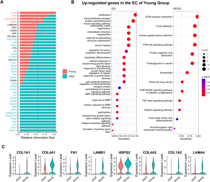
Background: Dental pulp regeneration therapy is a challenge to achieve early vascularization during treatment. Studying the regulatory mechanisms of vascular formation during human dental pulp development may provide insights for related therapies. In this study, we utilized single-cell sequencing analysis to compare the gene expression of dental pulp stem cells (DPSCs) and vascular endothelial cells (ECs) from developing and mature dental pulps.
Method: Immunohistochemistry, Western blot, and real-time polymerase chain reaction (RT-PCR) were used to detect fibronectin 1 (FN1) expression and molecules, such as PI3K/AKT. Cell proliferation assay, scratch assay, tube formation assay and were used to investigate the effects of DPSCs on the vasculogenetic capability of ECs. Additionally, animal experiments involving mice were conducted.
Result: The results revealed that DPSCs exist around dental pulp vasculature. FN1 expression was significantly higher in DPSCs from young permanent pulps than mature pulps, promoting HUVEC proliferation, migration, and tube formation via ITGA5 and the downstream PI3K/AKT signaling pathway.
Conclusion: Our data indicate that intercellular communication between DPSCs and ECs mediated by FN1-ITGA5 signaling is crucial for vascularizationduring dental pulp development, laying an experimental foundation for future clinical studies.
期刊介绍:
The purpose of Stem Cell Reviews and Reports is to cover contemporary and emerging areas in stem cell research and regenerative medicine. The journal will consider for publication:
i) solicited or unsolicited reviews of topical areas of stem cell biology that highlight, critique and synthesize recent important findings in the field.
ii) full length and short reports presenting original experimental work.
iii) translational stem cell studies describing results of clinical trials using stem cells as therapeutics.
iv) papers focused on diseases of stem cells.
v) hypothesis and commentary articles as opinion-based pieces in which authors can propose a new theory, interpretation of a controversial area in stem cell biology, or a stem cell biology question or paradigm. These articles contain more speculation than reviews, but they should be based on solid rationale.
vi) protocols as peer-reviewed procedures that provide step-by-step descriptions, outlined in sufficient detail, so that both experts and novices can apply them to their own research.
vii) letters to the editor and correspondence.
In order to facilitate this exchange of scientific information and exciting novel ideas, the journal has created five thematic sections, focusing on:
i) the role of adult stem cells in tissue regeneration;
ii) progress in research on induced pluripotent stem cells, embryonic stem cells and mechanism governing embryogenesis and tissue development;
iii) the role of microenvironment and extracellular microvesicles in directing the fate of stem cells;
iv) mechanisms of stem cell trafficking, stem cell mobilization and homing with special emphasis on hematopoiesis;
v) the role of stem cells in aging processes and cancerogenesis.


 分享
分享
 求助内容:
求助内容: 应助结果提醒方式:
 扫码关注我们
