下载PDF
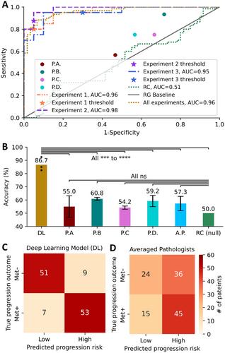
{"title":"人工智能引导的组织病理学可预测肺癌患者的脑转移。","authors":"Haowen Zhou, Mark Watson, Cory T Bernadt, Steven (Siyu) Lin, Chieh-yu Lin, Jon H Ritter, Alexander Wein, Simon Mahler, Sid Rawal, Ramaswamy Govindan, Changhuei Yang, Richard J Cote","doi":"10.1002/path.6263","DOIUrl":null,"url":null,"abstract":"<p>Brain metastases can occur in nearly half of patients with early and locally advanced (stage I–III) non-small cell lung cancer (NSCLC). There are no reliable histopathologic or molecular means to identify those who are likely to develop brain metastases. We sought to determine if deep learning (DL) could be applied to routine H&E-stained primary tumor tissue sections from stage I–III NSCLC patients to predict the development of brain metastasis. Diagnostic slides from 158 patients with stage I–III NSCLC followed for at least 5 years for the development of brain metastases (Met<sup>+</sup>, 65 patients) versus no progression (Met<sup>−</sup>, 93 patients) were subjected to whole-slide imaging. Three separate iterations were performed by first selecting 118 cases (45 Met<sup>+</sup>, 73 Met<sup>−</sup>) to train and validate the DL algorithm, while 40 separate cases (20 Met<sup>+</sup>, 20 Met<sup>−</sup>) were used as the test set. The DL algorithm results were compared to a blinded review by four expert pathologists. The DL-based algorithm was able to distinguish the eventual development of brain metastases with an accuracy of 87% (<i>p</i> < 0.0001) compared with an average of 57.3% by the four pathologists and appears to be particularly useful in predicting brain metastases in stage I patients. The DL algorithm appears to focus on a complex set of histologic features. DL-based algorithms using routine H&E-stained slides may identify patients who are likely to develop brain metastases from those who will remain disease free over extended (>5 year) follow-up and may thus be spared systemic therapy. © 2024 The Authors. <i>The Journal of Pathology</i> published by John Wiley & Sons Ltd on behalf of The Pathological Society of Great Britain and Ireland.</p>","PeriodicalId":232,"journal":{"name":"The Journal of Pathology","volume":"263 1","pages":"89-98"},"PeriodicalIF":5.6000,"publicationDate":"2024-03-04","publicationTypes":"Journal Article","fieldsOfStudy":null,"isOpenAccess":false,"openAccessPdf":"https://onlinelibrary.wiley.com/doi/epdf/10.1002/path.6263","citationCount":"0","resultStr":"{\"title\":\"AI-guided histopathology predicts brain metastasis in lung cancer patients\",\"authors\":\"Haowen Zhou, Mark Watson, Cory T Bernadt, Steven (Siyu) Lin, Chieh-yu Lin, Jon H Ritter, Alexander Wein, Simon Mahler, Sid Rawal, Ramaswamy Govindan, Changhuei Yang, Richard J Cote\",\"doi\":\"10.1002/path.6263\",\"DOIUrl\":null,\"url\":null,\"abstract\":\"<p>Brain metastases can occur in nearly half of patients with early and locally advanced (stage I–III) non-small cell lung cancer (NSCLC). There are no reliable histopathologic or molecular means to identify those who are likely to develop brain metastases. We sought to determine if deep learning (DL) could be applied to routine H&E-stained primary tumor tissue sections from stage I–III NSCLC patients to predict the development of brain metastasis. Diagnostic slides from 158 patients with stage I–III NSCLC followed for at least 5 years for the development of brain metastases (Met<sup>+</sup>, 65 patients) versus no progression (Met<sup>−</sup>, 93 patients) were subjected to whole-slide imaging. Three separate iterations were performed by first selecting 118 cases (45 Met<sup>+</sup>, 73 Met<sup>−</sup>) to train and validate the DL algorithm, while 40 separate cases (20 Met<sup>+</sup>, 20 Met<sup>−</sup>) were used as the test set. The DL algorithm results were compared to a blinded review by four expert pathologists. The DL-based algorithm was able to distinguish the eventual development of brain metastases with an accuracy of 87% (<i>p</i> < 0.0001) compared with an average of 57.3% by the four pathologists and appears to be particularly useful in predicting brain metastases in stage I patients. The DL algorithm appears to focus on a complex set of histologic features. DL-based algorithms using routine H&E-stained slides may identify patients who are likely to develop brain metastases from those who will remain disease free over extended (>5 year) follow-up and may thus be spared systemic therapy. © 2024 The Authors. <i>The Journal of Pathology</i> published by John Wiley & Sons Ltd on behalf of The Pathological Society of Great Britain and Ireland.</p>\",\"PeriodicalId\":232,\"journal\":{\"name\":\"The Journal of Pathology\",\"volume\":\"263 1\",\"pages\":\"89-98\"},\"PeriodicalIF\":5.6000,\"publicationDate\":\"2024-03-04\",\"publicationTypes\":\"Journal Article\",\"fieldsOfStudy\":null,\"isOpenAccess\":false,\"openAccessPdf\":\"https://onlinelibrary.wiley.com/doi/epdf/10.1002/path.6263\",\"citationCount\":\"0\",\"resultStr\":null,\"platform\":\"Semanticscholar\",\"paperid\":null,\"PeriodicalName\":\"The Journal of Pathology\",\"FirstCategoryId\":\"3\",\"ListUrlMain\":\"https://onlinelibrary.wiley.com/doi/10.1002/path.6263\",\"RegionNum\":2,\"RegionCategory\":\"医学\",\"ArticlePicture\":[],\"TitleCN\":null,\"AbstractTextCN\":null,\"PMCID\":null,\"EPubDate\":\"\",\"PubModel\":\"\",\"JCR\":\"Q1\",\"JCRName\":\"ONCOLOGY\",\"Score\":null,\"Total\":0}","platform":"Semanticscholar","paperid":null,"PeriodicalName":"The Journal of Pathology","FirstCategoryId":"3","ListUrlMain":"https://onlinelibrary.wiley.com/doi/10.1002/path.6263","RegionNum":2,"RegionCategory":"医学","ArticlePicture":[],"TitleCN":null,"AbstractTextCN":null,"PMCID":null,"EPubDate":"","PubModel":"","JCR":"Q1","JCRName":"ONCOLOGY","Score":null,"Total":0}
引用次数: 0
引用
批量引用


