{"title":"颅骨骨内血管畸形:病例报告和文献综述。","authors":"Donghyun Lee, Chul Hoon Chung, Seong Jin Cho","doi":"10.7181/acfs.2023.00584","DOIUrl":null,"url":null,"abstract":"<p><p>A 59-year-old woman presented to our clinic with a 3.5× 3-cm protruding mass on her forehead. A skull X-ray revealed a radiolucent osteolytic lesion on the left side of the frontal bone. Additionally, computed tomography showed a 3.1× 1.7× 3.6-cm mass exhibiting a \"sunburst\" pattern situated between the outer and inner tables of the skull, just superior and lateral to the left frontal sinus. This pattern suggested the presence of an intraosseous vascular malformation (IVM). The lesion was approached via a bicoronal incision. En-bloc resection was performed, removing the mass along with approximately 0.5 cm of the surrounding normal bone without injury to the exposed frontal sinus mucosa. The exposed mucosa was reinforced with a galeal flap, and cranioplasty with bone cement was performed to repair the resulting bony defect. Pathological examination confirmed a diagnosis of intraosseous cavernous-type malformation with mixed cavernous and capillary histological features. We report this case of IVM and review the existing literature, highlighting the satisfactory functional and aesthetic outcomes after surgery.</p>","PeriodicalId":52238,"journal":{"name":"Archives of Craniofacial Surgery","volume":" ","pages":"187-191"},"PeriodicalIF":0.0000,"publicationDate":"2024-08-01","publicationTypes":"Journal Article","fieldsOfStudy":null,"isOpenAccess":false,"openAccessPdf":"https://www.ncbi.nlm.nih.gov/pmc/articles/PMC11374518/pdf/","citationCount":"0","resultStr":"{\"title\":\"Intraosseous vascular malformation of the skull: a case report and literature review.\",\"authors\":\"Donghyun Lee, Chul Hoon Chung, Seong Jin Cho\",\"doi\":\"10.7181/acfs.2023.00584\",\"DOIUrl\":null,\"url\":null,\"abstract\":\"<p><p>A 59-year-old woman presented to our clinic with a 3.5× 3-cm protruding mass on her forehead. A skull X-ray revealed a radiolucent osteolytic lesion on the left side of the frontal bone. Additionally, computed tomography showed a 3.1× 1.7× 3.6-cm mass exhibiting a \\\"sunburst\\\" pattern situated between the outer and inner tables of the skull, just superior and lateral to the left frontal sinus. This pattern suggested the presence of an intraosseous vascular malformation (IVM). The lesion was approached via a bicoronal incision. En-bloc resection was performed, removing the mass along with approximately 0.5 cm of the surrounding normal bone without injury to the exposed frontal sinus mucosa. The exposed mucosa was reinforced with a galeal flap, and cranioplasty with bone cement was performed to repair the resulting bony defect. Pathological examination confirmed a diagnosis of intraosseous cavernous-type malformation with mixed cavernous and capillary histological features. We report this case of IVM and review the existing literature, highlighting the satisfactory functional and aesthetic outcomes after surgery.</p>\",\"PeriodicalId\":52238,\"journal\":{\"name\":\"Archives of Craniofacial Surgery\",\"volume\":\" \",\"pages\":\"187-191\"},\"PeriodicalIF\":0.0000,\"publicationDate\":\"2024-08-01\",\"publicationTypes\":\"Journal Article\",\"fieldsOfStudy\":null,\"isOpenAccess\":false,\"openAccessPdf\":\"https://www.ncbi.nlm.nih.gov/pmc/articles/PMC11374518/pdf/\",\"citationCount\":\"0\",\"resultStr\":null,\"platform\":\"Semanticscholar\",\"paperid\":null,\"PeriodicalName\":\"Archives of Craniofacial Surgery\",\"FirstCategoryId\":\"1085\",\"ListUrlMain\":\"https://doi.org/10.7181/acfs.2023.00584\",\"RegionNum\":0,\"RegionCategory\":null,\"ArticlePicture\":[],\"TitleCN\":null,\"AbstractTextCN\":null,\"PMCID\":null,\"EPubDate\":\"2024/3/6 0:00:00\",\"PubModel\":\"Epub\",\"JCR\":\"Q2\",\"JCRName\":\"Medicine\",\"Score\":null,\"Total\":0}","platform":"Semanticscholar","paperid":null,"PeriodicalName":"Archives of Craniofacial Surgery","FirstCategoryId":"1085","ListUrlMain":"https://doi.org/10.7181/acfs.2023.00584","RegionNum":0,"RegionCategory":null,"ArticlePicture":[],"TitleCN":null,"AbstractTextCN":null,"PMCID":null,"EPubDate":"2024/3/6 0:00:00","PubModel":"Epub","JCR":"Q2","JCRName":"Medicine","Score":null,"Total":0}
Intraosseous vascular malformation of the skull: a case report and literature review.
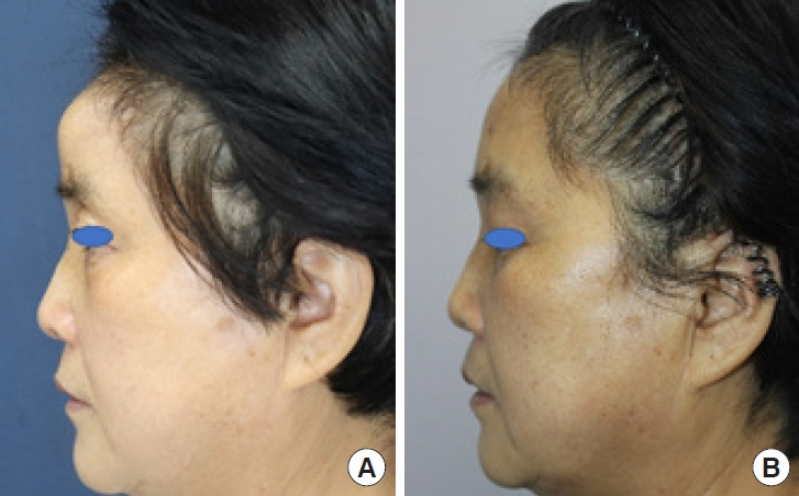
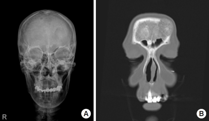
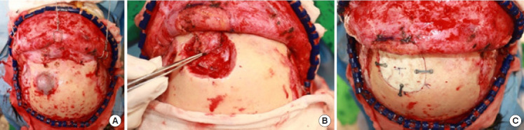
A 59-year-old woman presented to our clinic with a 3.5× 3-cm protruding mass on her forehead. A skull X-ray revealed a radiolucent osteolytic lesion on the left side of the frontal bone. Additionally, computed tomography showed a 3.1× 1.7× 3.6-cm mass exhibiting a "sunburst" pattern situated between the outer and inner tables of the skull, just superior and lateral to the left frontal sinus. This pattern suggested the presence of an intraosseous vascular malformation (IVM). The lesion was approached via a bicoronal incision. En-bloc resection was performed, removing the mass along with approximately 0.5 cm of the surrounding normal bone without injury to the exposed frontal sinus mucosa. The exposed mucosa was reinforced with a galeal flap, and cranioplasty with bone cement was performed to repair the resulting bony defect. Pathological examination confirmed a diagnosis of intraosseous cavernous-type malformation with mixed cavernous and capillary histological features. We report this case of IVM and review the existing literature, highlighting the satisfactory functional and aesthetic outcomes after surgery.


 分享
分享
 求助内容:
求助内容: 应助结果提醒方式:
 扫码关注我们
