{"title":"中肾样子宫内膜腺癌的子宫内膜细胞学发现:病例报告。","authors":"Hirotaka Omine CT, Kazuyuki Ishida MD, PhD, Natsuki Sasaki CT, Hikaru Kato CT, Tamiko Nagai CT, Mihoko Ishikawa CT, Mina Takaoka MD, Shuhei Noda MD, Hadzki Matsuda MD, PhD, Akira Mitsuhashi MD, PhD","doi":"10.1002/dc.25300","DOIUrl":null,"url":null,"abstract":"<p>A mesonephric-like endometrial adenocarcinoma (ML-EAC) is very rare and has a worse prognosis than other endometrial carcinomas. We describe an ML-EAC and report our endometrial cytological findings. A 76-year-old woman presented with irregular genital bleeding and a uterine mass. Endometrial cytology revealed atypical cylindrical or spindle-shaped cells in the form of small aggregates or solitary cells. The cell aggregates exhibited irregularly stacked papillary structures, small glandular structures, and fenestrated structures. The atypical cells had a nucleus with fine-granular chromatin and a granular cytoplasm, and nuclear grooves and intranuclear pseudo-inclusions were present. Hyaline globules were observed in the glandular lumens and in the background. The presumptive histological type was an adenocarcinoma, but the cytological features were different from those of an endometrioid carcinoma. A histological examination of the endometrial biopsy revealed an adenocarcinoma, and a simple hysterectomy was performed. A grayish-white elevated mass measuring 90 mm × 70 mm × 40 mm was observed on the uterine corpus in the hysterectomy specimen. Histologically, the tumor proliferated as complex tubular structures containing eosinophilic colloid-like materials and trabecular structures. The tumor cells were diffuse and positive for GATA-3 and partially positive for thyroid transcription factor-1. Estrogen and progesterone receptors were negative. An ML-EAC was diagnosed. The tumor was invasive and extended beyond one-half of the muscle layer with a high degree of vascular invasion. In conclusion, we need to focus on the various shapes of the cell aggregate, nuclear grooves, and intranuclear pseudo-inclusions of tumor cells to distinguish an ML-EAC from other endometrial carcinomas in endometrial cytology.</p>","PeriodicalId":11349,"journal":{"name":"Diagnostic Cytopathology","volume":"52 6","pages":"E129-E133"},"PeriodicalIF":0.9000,"publicationDate":"2024-03-07","publicationTypes":"Journal Article","fieldsOfStudy":null,"isOpenAccess":false,"openAccessPdf":"https://onlinelibrary.wiley.com/doi/epdf/10.1002/dc.25300","citationCount":"0","resultStr":"{\"title\":\"Endometrial cytological findings for a mesonephric-like endometrial adenocarcinoma: A case report\",\"authors\":\"Hirotaka Omine CT, Kazuyuki Ishida MD, PhD, Natsuki Sasaki CT, Hikaru Kato CT, Tamiko Nagai CT, Mihoko Ishikawa CT, Mina Takaoka MD, Shuhei Noda MD, Hadzki Matsuda MD, PhD, Akira Mitsuhashi MD, PhD\",\"doi\":\"10.1002/dc.25300\",\"DOIUrl\":null,\"url\":null,\"abstract\":\"<p>A mesonephric-like endometrial adenocarcinoma (ML-EAC) is very rare and has a worse prognosis than other endometrial carcinomas. We describe an ML-EAC and report our endometrial cytological findings. A 76-year-old woman presented with irregular genital bleeding and a uterine mass. Endometrial cytology revealed atypical cylindrical or spindle-shaped cells in the form of small aggregates or solitary cells. The cell aggregates exhibited irregularly stacked papillary structures, small glandular structures, and fenestrated structures. The atypical cells had a nucleus with fine-granular chromatin and a granular cytoplasm, and nuclear grooves and intranuclear pseudo-inclusions were present. Hyaline globules were observed in the glandular lumens and in the background. The presumptive histological type was an adenocarcinoma, but the cytological features were different from those of an endometrioid carcinoma. A histological examination of the endometrial biopsy revealed an adenocarcinoma, and a simple hysterectomy was performed. A grayish-white elevated mass measuring 90 mm × 70 mm × 40 mm was observed on the uterine corpus in the hysterectomy specimen. Histologically, the tumor proliferated as complex tubular structures containing eosinophilic colloid-like materials and trabecular structures. The tumor cells were diffuse and positive for GATA-3 and partially positive for thyroid transcription factor-1. Estrogen and progesterone receptors were negative. An ML-EAC was diagnosed. The tumor was invasive and extended beyond one-half of the muscle layer with a high degree of vascular invasion. In conclusion, we need to focus on the various shapes of the cell aggregate, nuclear grooves, and intranuclear pseudo-inclusions of tumor cells to distinguish an ML-EAC from other endometrial carcinomas in endometrial cytology.</p>\",\"PeriodicalId\":11349,\"journal\":{\"name\":\"Diagnostic Cytopathology\",\"volume\":\"52 6\",\"pages\":\"E129-E133\"},\"PeriodicalIF\":0.9000,\"publicationDate\":\"2024-03-07\",\"publicationTypes\":\"Journal Article\",\"fieldsOfStudy\":null,\"isOpenAccess\":false,\"openAccessPdf\":\"https://onlinelibrary.wiley.com/doi/epdf/10.1002/dc.25300\",\"citationCount\":\"0\",\"resultStr\":null,\"platform\":\"Semanticscholar\",\"paperid\":null,\"PeriodicalName\":\"Diagnostic Cytopathology\",\"FirstCategoryId\":\"3\",\"ListUrlMain\":\"https://onlinelibrary.wiley.com/doi/10.1002/dc.25300\",\"RegionNum\":4,\"RegionCategory\":\"医学\",\"ArticlePicture\":[],\"TitleCN\":null,\"AbstractTextCN\":null,\"PMCID\":null,\"EPubDate\":\"\",\"PubModel\":\"\",\"JCR\":\"Q4\",\"JCRName\":\"MEDICAL LABORATORY TECHNOLOGY\",\"Score\":null,\"Total\":0}","platform":"Semanticscholar","paperid":null,"PeriodicalName":"Diagnostic Cytopathology","FirstCategoryId":"3","ListUrlMain":"https://onlinelibrary.wiley.com/doi/10.1002/dc.25300","RegionNum":4,"RegionCategory":"医学","ArticlePicture":[],"TitleCN":null,"AbstractTextCN":null,"PMCID":null,"EPubDate":"","PubModel":"","JCR":"Q4","JCRName":"MEDICAL LABORATORY TECHNOLOGY","Score":null,"Total":0}
Endometrial cytological findings for a mesonephric-like endometrial adenocarcinoma: A case report
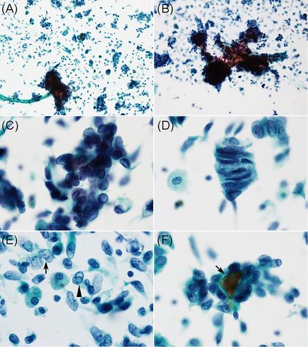
A mesonephric-like endometrial adenocarcinoma (ML-EAC) is very rare and has a worse prognosis than other endometrial carcinomas. We describe an ML-EAC and report our endometrial cytological findings. A 76-year-old woman presented with irregular genital bleeding and a uterine mass. Endometrial cytology revealed atypical cylindrical or spindle-shaped cells in the form of small aggregates or solitary cells. The cell aggregates exhibited irregularly stacked papillary structures, small glandular structures, and fenestrated structures. The atypical cells had a nucleus with fine-granular chromatin and a granular cytoplasm, and nuclear grooves and intranuclear pseudo-inclusions were present. Hyaline globules were observed in the glandular lumens and in the background. The presumptive histological type was an adenocarcinoma, but the cytological features were different from those of an endometrioid carcinoma. A histological examination of the endometrial biopsy revealed an adenocarcinoma, and a simple hysterectomy was performed. A grayish-white elevated mass measuring 90 mm × 70 mm × 40 mm was observed on the uterine corpus in the hysterectomy specimen. Histologically, the tumor proliferated as complex tubular structures containing eosinophilic colloid-like materials and trabecular structures. The tumor cells were diffuse and positive for GATA-3 and partially positive for thyroid transcription factor-1. Estrogen and progesterone receptors were negative. An ML-EAC was diagnosed. The tumor was invasive and extended beyond one-half of the muscle layer with a high degree of vascular invasion. In conclusion, we need to focus on the various shapes of the cell aggregate, nuclear grooves, and intranuclear pseudo-inclusions of tumor cells to distinguish an ML-EAC from other endometrial carcinomas in endometrial cytology.
期刊介绍:
Diagnostic Cytopathology is intended to provide a forum for the exchange of information in the field of cytopathology, with special emphasis on the practical, clinical aspects of the discipline. The editors invite original scientific articles, as well as special review articles, feature articles, and letters to the editor, from laboratory professionals engaged in the practice of cytopathology. Manuscripts are accepted for publication on the basis of scientific merit, practical significance, and suitability for publication in a journal dedicated to this discipline. Original articles can be considered only with the understanding that they have never been published before and that they have not been submitted for simultaneous review to another publication.


 分享
分享
 求助内容:
求助内容: 应助结果提醒方式:
 扫码关注我们
