Yunus Imren, Bulent Karslioğlu, Suleyman Semih Dedeoğlu, Ahmet Keskin, Ahmet Firat Berkay, Ali Cagri Tekin
{"title":"利用髋部骨折常规计算机断层扫描诊断骨质疏松症的可行性:与头颈部组织病理学诊断的相关性。","authors":"Yunus Imren, Bulent Karslioğlu, Suleyman Semih Dedeoğlu, Ahmet Keskin, Ahmet Firat Berkay, Ali Cagri Tekin","doi":"10.5152/j.aott.2023.23126","DOIUrl":null,"url":null,"abstract":"<p><strong>Objective: </strong>The aim of this study was to demonstrate the feasibility of diagnosing osteoporosis through routine computed tomography (CT) by assessing the association between the histopathological assessment of femoral head specimens extracted from patients who underwent surgery for intertrochanteric fractures and the Hounsfield unit (HU) measurements derived from preoperative CT scans.</p><p><strong>Methods: </strong>Forty-eight patients who presented to our clinic between November 2019 and May 2020 with hip fractures and underwent partial prosthesis fixation were included in this retrospective study. Hounsfield unit measurements were performed on the head and neck regions using dual-energy x-ray absorptiometry (DEXA) and CT scans, respectively. The trabecular ratio per unit area was calculated using the Nikon Imaging Software (NIS-Elements ) program in the pathology laboratory from digitally captured images of the removed head and neck specimens.</p><p><strong>Results: </strong>The mean HU receiver operating characteristic analysis had a sensitivity of 77% and a specificity of 87%, with a cutoff value of 77.68. There was a moderate correlation between the mean trabecular density and the mean HU of the femoral head (P=0.013, r=0.340). Additionally, there was a significant correlation between the mean HU and the T-score of the head, although this correlation was not found with the maximum-minimum HU. Although there was a significant correlation between trabecular density and mean HU, the correlation coefficient indicated a moderate relationship. This relationship was also observed between the inferior sections of the head and the trabecular density and HU (P=.018). However, no significant correlation was found between the T-score and the trabecular structure of the head (P=.977).</p><p><strong>Conclusion: </strong>The results of the present study suggest that conventional CT has the potential to serve as a diagnostic tool for osteoporosis and may offer a more precise and accurate method for evaluating the success of intraosseous implants when compared to T-scores without the need for additional tests or procedures.</p>","PeriodicalId":93854,"journal":{"name":"Acta orthopaedica et traumatologica turcica","volume":"57 6","pages":"384-388"},"PeriodicalIF":1.0000,"publicationDate":"2023-11-01","publicationTypes":"Journal Article","fieldsOfStudy":null,"isOpenAccess":false,"openAccessPdf":"https://www.ncbi.nlm.nih.gov/pmc/articles/PMC10837590/pdf/","citationCount":"0","resultStr":"{\"title\":\"Feasibility of diagnosing osteoporosis using routine computed tomography scans for hip fractures: Correlation with histopathological diagnosis of head and neck regions.\",\"authors\":\"Yunus Imren, Bulent Karslioğlu, Suleyman Semih Dedeoğlu, Ahmet Keskin, Ahmet Firat Berkay, Ali Cagri Tekin\",\"doi\":\"10.5152/j.aott.2023.23126\",\"DOIUrl\":null,\"url\":null,\"abstract\":\"<p><strong>Objective: </strong>The aim of this study was to demonstrate the feasibility of diagnosing osteoporosis through routine computed tomography (CT) by assessing the association between the histopathological assessment of femoral head specimens extracted from patients who underwent surgery for intertrochanteric fractures and the Hounsfield unit (HU) measurements derived from preoperative CT scans.</p><p><strong>Methods: </strong>Forty-eight patients who presented to our clinic between November 2019 and May 2020 with hip fractures and underwent partial prosthesis fixation were included in this retrospective study. Hounsfield unit measurements were performed on the head and neck regions using dual-energy x-ray absorptiometry (DEXA) and CT scans, respectively. The trabecular ratio per unit area was calculated using the Nikon Imaging Software (NIS-Elements ) program in the pathology laboratory from digitally captured images of the removed head and neck specimens.</p><p><strong>Results: </strong>The mean HU receiver operating characteristic analysis had a sensitivity of 77% and a specificity of 87%, with a cutoff value of 77.68. There was a moderate correlation between the mean trabecular density and the mean HU of the femoral head (P=0.013, r=0.340). Additionally, there was a significant correlation between the mean HU and the T-score of the head, although this correlation was not found with the maximum-minimum HU. Although there was a significant correlation between trabecular density and mean HU, the correlation coefficient indicated a moderate relationship. This relationship was also observed between the inferior sections of the head and the trabecular density and HU (P=.018). However, no significant correlation was found between the T-score and the trabecular structure of the head (P=.977).</p><p><strong>Conclusion: </strong>The results of the present study suggest that conventional CT has the potential to serve as a diagnostic tool for osteoporosis and may offer a more precise and accurate method for evaluating the success of intraosseous implants when compared to T-scores without the need for additional tests or procedures.</p>\",\"PeriodicalId\":93854,\"journal\":{\"name\":\"Acta orthopaedica et traumatologica turcica\",\"volume\":\"57 6\",\"pages\":\"384-388\"},\"PeriodicalIF\":1.0000,\"publicationDate\":\"2023-11-01\",\"publicationTypes\":\"Journal Article\",\"fieldsOfStudy\":null,\"isOpenAccess\":false,\"openAccessPdf\":\"https://www.ncbi.nlm.nih.gov/pmc/articles/PMC10837590/pdf/\",\"citationCount\":\"0\",\"resultStr\":null,\"platform\":\"Semanticscholar\",\"paperid\":null,\"PeriodicalName\":\"Acta orthopaedica et traumatologica turcica\",\"FirstCategoryId\":\"1085\",\"ListUrlMain\":\"https://doi.org/10.5152/j.aott.2023.23126\",\"RegionNum\":0,\"RegionCategory\":null,\"ArticlePicture\":[],\"TitleCN\":null,\"AbstractTextCN\":null,\"PMCID\":null,\"EPubDate\":\"\",\"PubModel\":\"\",\"JCR\":\"\",\"JCRName\":\"\",\"Score\":null,\"Total\":0}","platform":"Semanticscholar","paperid":null,"PeriodicalName":"Acta orthopaedica et traumatologica turcica","FirstCategoryId":"1085","ListUrlMain":"https://doi.org/10.5152/j.aott.2023.23126","RegionNum":0,"RegionCategory":null,"ArticlePicture":[],"TitleCN":null,"AbstractTextCN":null,"PMCID":null,"EPubDate":"","PubModel":"","JCR":"","JCRName":"","Score":null,"Total":0}
引用次数: 0
摘要
研究目的本研究旨在通过评估从接受转子间骨折手术的患者身上提取的股骨头标本的组织病理学评估与术前 CT 扫描得出的 Hounsfield 单位(HU)测量值之间的关联,证明通过常规计算机断层扫描(CT)诊断骨质疏松症的可行性:本回顾性研究共纳入了 48 名在 2019 年 11 月至 2020 年 5 月期间因髋部骨折就诊并接受部分假体固定术的患者。分别使用双能 X 射线吸收测量法(DEXA)和 CT 扫描对患者的头部和颈部进行 Hounsfield 单位测量。病理实验室使用尼康成像软件(NIS-Elements)程序,从切除的头颈部标本的数字采集图像中计算出单位面积的小梁比率:平均 HU 接受者操作特征分析的灵敏度为 77%,特异度为 87%,临界值为 77.68。股骨头的平均骨小梁密度与平均 HU 之间存在中度相关性(P=0.013,r=0.340)。此外,平均 HU 值与股骨头的 T 评分之间也存在显著相关性,但最大-最小 HU 值之间则不存在相关性。虽然小梁密度与平均 HU 之间存在明显的相关性,但相关系数仅为中等水平。在头部下段与小梁密度和 HU 之间也观察到这种关系(P=.018)。然而,T 评分与头部小梁结构之间没有发现明显的相关性(P=.977):本研究结果表明,常规 CT 有潜力成为骨质疏松症的诊断工具,与 T 评分相比,它可以提供一种更精确、更准确的方法来评估骨内植入物的成功率,而无需额外的测试或程序。
Feasibility of diagnosing osteoporosis using routine computed tomography scans for hip fractures: Correlation with histopathological diagnosis of head and neck regions.
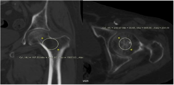
Objective: The aim of this study was to demonstrate the feasibility of diagnosing osteoporosis through routine computed tomography (CT) by assessing the association between the histopathological assessment of femoral head specimens extracted from patients who underwent surgery for intertrochanteric fractures and the Hounsfield unit (HU) measurements derived from preoperative CT scans.
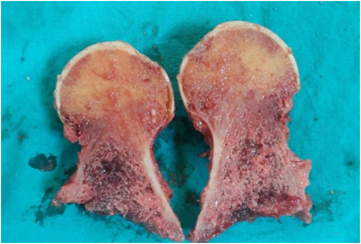
Methods: Forty-eight patients who presented to our clinic between November 2019 and May 2020 with hip fractures and underwent partial prosthesis fixation were included in this retrospective study. Hounsfield unit measurements were performed on the head and neck regions using dual-energy x-ray absorptiometry (DEXA) and CT scans, respectively. The trabecular ratio per unit area was calculated using the Nikon Imaging Software (NIS-Elements ) program in the pathology laboratory from digitally captured images of the removed head and neck specimens.
Results: The mean HU receiver operating characteristic analysis had a sensitivity of 77% and a specificity of 87%, with a cutoff value of 77.68. There was a moderate correlation between the mean trabecular density and the mean HU of the femoral head (P=0.013, r=0.340). Additionally, there was a significant correlation between the mean HU and the T-score of the head, although this correlation was not found with the maximum-minimum HU. Although there was a significant correlation between trabecular density and mean HU, the correlation coefficient indicated a moderate relationship. This relationship was also observed between the inferior sections of the head and the trabecular density and HU (P=.018). However, no significant correlation was found between the T-score and the trabecular structure of the head (P=.977).
Conclusion: The results of the present study suggest that conventional CT has the potential to serve as a diagnostic tool for osteoporosis and may offer a more precise and accurate method for evaluating the success of intraosseous implants when compared to T-scores without the need for additional tests or procedures.


 分享
分享

 求助内容:
求助内容: 应助结果提醒方式:
 扫码关注我们
