Ahmed Shah MD, MSc, Adrian Box MD, PhD, Thomas Brenn MD, PhD, Ashley Flaman MD
{"title":"原发性皮肤 NUT 癌,伴有 BRD4::NUTM1 融合。","authors":"Ahmed Shah MD, MSc, Adrian Box MD, PhD, Thomas Brenn MD, PhD, Ashley Flaman MD","doi":"10.1111/cup.14602","DOIUrl":null,"url":null,"abstract":"<p>Nuclear protein in testis (NUT) carcinoma, molecularly defined by the <i>NUTM1</i> gene rearrangement, is most commonly reported in young adults in the sinonasal tract, nasopharynx, or thorax. At these sites, NUT carcinoma is an extremely aggressive malignancy with dismal prognosis. Recently, five cases of primary cutaneous NUT adnexal carcinoma have been reported with <i>BRD3</i> and <i>NSD3</i> fusion partners. Although NUT adnexal carcinomas are shown to have metastatic potential, they may behave less aggressively than extracutaneous NUT carcinomas. We report a case of a 59-year-old man who underwent a biopsy of a 3-cm plantar mass, which showed <i>BRD4::NUTM1</i> fusion. The tumor was a poorly differentiated dermal neoplasm showing cytologic atypia, large vesicular nuclei with prominent nucleoli, conspicuous mitotic activity, and foci of necrosis. Immunohistochemically, the tumor showed positivity for keratins, EMA, SOX10, and NUT, with patchy smooth muscle actin. Molecular testing revealed <i>BRD4::NUTM1</i> rearrangement. With no alternative primary identified by imaging, a diagnosis of primary cutaneous NUT carcinoma was favored. We hope to contribute to the limited body of knowledge on this entity, with emphasis on recognition as well as studying and defining its prognostic differences from extracutaneous NUT carcinomas.</p>","PeriodicalId":15407,"journal":{"name":"Journal of Cutaneous Pathology","volume":"51 6","pages":"424-429"},"PeriodicalIF":1.1000,"publicationDate":"2024-03-13","publicationTypes":"Journal Article","fieldsOfStudy":null,"isOpenAccess":false,"openAccessPdf":"https://onlinelibrary.wiley.com/doi/epdf/10.1111/cup.14602","citationCount":"0","resultStr":"{\"title\":\"Primary cutaneous NUT carcinoma with BRD4::NUTM1 fusion\",\"authors\":\"Ahmed Shah MD, MSc, Adrian Box MD, PhD, Thomas Brenn MD, PhD, Ashley Flaman MD\",\"doi\":\"10.1111/cup.14602\",\"DOIUrl\":null,\"url\":null,\"abstract\":\"<p>Nuclear protein in testis (NUT) carcinoma, molecularly defined by the <i>NUTM1</i> gene rearrangement, is most commonly reported in young adults in the sinonasal tract, nasopharynx, or thorax. At these sites, NUT carcinoma is an extremely aggressive malignancy with dismal prognosis. Recently, five cases of primary cutaneous NUT adnexal carcinoma have been reported with <i>BRD3</i> and <i>NSD3</i> fusion partners. Although NUT adnexal carcinomas are shown to have metastatic potential, they may behave less aggressively than extracutaneous NUT carcinomas. We report a case of a 59-year-old man who underwent a biopsy of a 3-cm plantar mass, which showed <i>BRD4::NUTM1</i> fusion. The tumor was a poorly differentiated dermal neoplasm showing cytologic atypia, large vesicular nuclei with prominent nucleoli, conspicuous mitotic activity, and foci of necrosis. Immunohistochemically, the tumor showed positivity for keratins, EMA, SOX10, and NUT, with patchy smooth muscle actin. Molecular testing revealed <i>BRD4::NUTM1</i> rearrangement. With no alternative primary identified by imaging, a diagnosis of primary cutaneous NUT carcinoma was favored. We hope to contribute to the limited body of knowledge on this entity, with emphasis on recognition as well as studying and defining its prognostic differences from extracutaneous NUT carcinomas.</p>\",\"PeriodicalId\":15407,\"journal\":{\"name\":\"Journal of Cutaneous Pathology\",\"volume\":\"51 6\",\"pages\":\"424-429\"},\"PeriodicalIF\":1.1000,\"publicationDate\":\"2024-03-13\",\"publicationTypes\":\"Journal Article\",\"fieldsOfStudy\":null,\"isOpenAccess\":false,\"openAccessPdf\":\"https://onlinelibrary.wiley.com/doi/epdf/10.1111/cup.14602\",\"citationCount\":\"0\",\"resultStr\":null,\"platform\":\"Semanticscholar\",\"paperid\":null,\"PeriodicalName\":\"Journal of Cutaneous Pathology\",\"FirstCategoryId\":\"3\",\"ListUrlMain\":\"https://onlinelibrary.wiley.com/doi/10.1111/cup.14602\",\"RegionNum\":4,\"RegionCategory\":\"医学\",\"ArticlePicture\":[],\"TitleCN\":null,\"AbstractTextCN\":null,\"PMCID\":null,\"EPubDate\":\"\",\"PubModel\":\"\",\"JCR\":\"Q3\",\"JCRName\":\"DERMATOLOGY\",\"Score\":null,\"Total\":0}","platform":"Semanticscholar","paperid":null,"PeriodicalName":"Journal of Cutaneous Pathology","FirstCategoryId":"3","ListUrlMain":"https://onlinelibrary.wiley.com/doi/10.1111/cup.14602","RegionNum":4,"RegionCategory":"医学","ArticlePicture":[],"TitleCN":null,"AbstractTextCN":null,"PMCID":null,"EPubDate":"","PubModel":"","JCR":"Q3","JCRName":"DERMATOLOGY","Score":null,"Total":0}
Primary cutaneous NUT carcinoma with BRD4::NUTM1 fusion
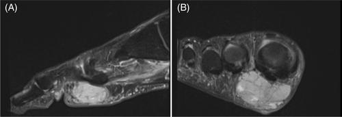
Nuclear protein in testis (NUT) carcinoma, molecularly defined by the NUTM1 gene rearrangement, is most commonly reported in young adults in the sinonasal tract, nasopharynx, or thorax. At these sites, NUT carcinoma is an extremely aggressive malignancy with dismal prognosis. Recently, five cases of primary cutaneous NUT adnexal carcinoma have been reported with BRD3 and NSD3 fusion partners. Although NUT adnexal carcinomas are shown to have metastatic potential, they may behave less aggressively than extracutaneous NUT carcinomas. We report a case of a 59-year-old man who underwent a biopsy of a 3-cm plantar mass, which showed BRD4::NUTM1 fusion. The tumor was a poorly differentiated dermal neoplasm showing cytologic atypia, large vesicular nuclei with prominent nucleoli, conspicuous mitotic activity, and foci of necrosis. Immunohistochemically, the tumor showed positivity for keratins, EMA, SOX10, and NUT, with patchy smooth muscle actin. Molecular testing revealed BRD4::NUTM1 rearrangement. With no alternative primary identified by imaging, a diagnosis of primary cutaneous NUT carcinoma was favored. We hope to contribute to the limited body of knowledge on this entity, with emphasis on recognition as well as studying and defining its prognostic differences from extracutaneous NUT carcinomas.
期刊介绍:
Journal of Cutaneous Pathology publishes manuscripts broadly relevant to diseases of the skin and mucosae, with the aims of advancing scientific knowledge regarding dermatopathology and enhancing the communication between clinical practitioners and research scientists. Original scientific manuscripts on diagnostic and experimental cutaneous pathology are especially desirable. Timely, pertinent review articles also will be given high priority. Manuscripts based on light, fluorescence, and electron microscopy, histochemistry, immunology, molecular biology, and genetics, as well as allied sciences, are all welcome, provided their principal focus is on cutaneous pathology. Publication time will be kept as short as possible, ensuring that articles will be quickly available to all interested in this speciality.


 分享
分享
 求助内容:
求助内容: 应助结果提醒方式:
 扫码关注我们
