{"title":"通过多参数神经元取向弥散和密度成像(NODDI)放射组学对胶质母细胞瘤和转移瘤进行高性能术前分化。","authors":"Jie Bai, Mengyang He, Eryuan Gao, Guang Yang, Chengxiu Zhang, Hongxi Yang, Jie Dong, Xiaoyue Ma, Yufei Gao, Huiting Zhang, Xu Yan, Yong Zhang, Jingliang Cheng, Guohua Zhao","doi":"10.1007/s00330-024-10686-8","DOIUrl":null,"url":null,"abstract":"<p><strong>Objectives: </strong>To evaluate the performance of multiparametric neurite orientation dispersion and density imaging (NODDI) radiomics in distinguishing between glioblastoma (Gb) and solitary brain metastasis (SBM).</p><p><strong>Materials and methods: </strong>In this retrospective study, NODDI images were curated from 109 patients with Gb (n = 57) or SBM (n = 52). Automatically segmented multiple volumes of interest (VOIs) encompassed the main tumor regions, including necrosis, solid tumor, and peritumoral edema. Radiomics features were extracted for each main tumor region, using three NODDI parameter maps. Radiomics models were developed based on these three NODDI parameter maps and their amalgamation to differentiate between Gb and SBM. Additionally, radiomics models were constructed based on morphological magnetic resonance imaging (MRI) and diffusion imaging (diffusion-weighted imaging [DWI]; diffusion tensor imaging [DTI]) for performance comparison.</p><p><strong>Results: </strong>The validation dataset results revealed that the performance of a single NODDI parameter map model was inferior to that of the combined NODDI model. In the necrotic regions, the combined NODDI radiomics model exhibited less than ideal discriminative capabilities (area under the receiver operating characteristic curve [AUC] = 0.701). For peritumoral edema regions, the combined NODDI radiomics model achieved a moderate level of discrimination (AUC = 0.820). Within the solid tumor regions, the combined NODDI radiomics model demonstrated superior performance (AUC = 0.904), surpassing the models of other VOIs. The comparison results demonstrated that the NODDI model was better than the DWI and DTI models, while those of the morphological MRI and NODDI models were similar.</p><p><strong>Conclusion: </strong>The NODDI radiomics model showed promising performance for preoperative discrimination between Gb and SBM.</p><p><strong>Clinical relevance statement: </strong>The NODDI radiomics model showed promising performance for preoperative discrimination between Gb and SBM, and radiomics features can be incorporated into the multidimensional phenotypic features that describe tumor heterogeneity.</p><p><strong>Key points: </strong>• The neurite orientation dispersion and density imaging (NODDI) radiomics model showed promising performance for preoperative discrimination between glioblastoma and solitary brain metastasis. • Compared with other tumor volumes of interest, the NODDI radiomics model based on solid tumor regions performed best in distinguishing the two types of tumors. • The performance of the single-parameter NODDI model was inferior to that of the combined-parameter NODDI model.</p>","PeriodicalId":12076,"journal":{"name":"European Radiology","volume":" ","pages":"6616-6628"},"PeriodicalIF":4.7000,"publicationDate":"2024-10-01","publicationTypes":"Journal Article","fieldsOfStudy":null,"isOpenAccess":false,"openAccessPdf":"https://www.ncbi.nlm.nih.gov/pmc/articles/PMC11399163/pdf/","citationCount":"0","resultStr":"{\"title\":\"High-performance presurgical differentiation of glioblastoma and metastasis by means of multiparametric neurite orientation dispersion and density imaging (NODDI) radiomics.\",\"authors\":\"Jie Bai, Mengyang He, Eryuan Gao, Guang Yang, Chengxiu Zhang, Hongxi Yang, Jie Dong, Xiaoyue Ma, Yufei Gao, Huiting Zhang, Xu Yan, Yong Zhang, Jingliang Cheng, Guohua Zhao\",\"doi\":\"10.1007/s00330-024-10686-8\",\"DOIUrl\":null,\"url\":null,\"abstract\":\"<p><strong>Objectives: </strong>To evaluate the performance of multiparametric neurite orientation dispersion and density imaging (NODDI) radiomics in distinguishing between glioblastoma (Gb) and solitary brain metastasis (SBM).</p><p><strong>Materials and methods: </strong>In this retrospective study, NODDI images were curated from 109 patients with Gb (n = 57) or SBM (n = 52). Automatically segmented multiple volumes of interest (VOIs) encompassed the main tumor regions, including necrosis, solid tumor, and peritumoral edema. Radiomics features were extracted for each main tumor region, using three NODDI parameter maps. Radiomics models were developed based on these three NODDI parameter maps and their amalgamation to differentiate between Gb and SBM. Additionally, radiomics models were constructed based on morphological magnetic resonance imaging (MRI) and diffusion imaging (diffusion-weighted imaging [DWI]; diffusion tensor imaging [DTI]) for performance comparison.</p><p><strong>Results: </strong>The validation dataset results revealed that the performance of a single NODDI parameter map model was inferior to that of the combined NODDI model. In the necrotic regions, the combined NODDI radiomics model exhibited less than ideal discriminative capabilities (area under the receiver operating characteristic curve [AUC] = 0.701). For peritumoral edema regions, the combined NODDI radiomics model achieved a moderate level of discrimination (AUC = 0.820). Within the solid tumor regions, the combined NODDI radiomics model demonstrated superior performance (AUC = 0.904), surpassing the models of other VOIs. The comparison results demonstrated that the NODDI model was better than the DWI and DTI models, while those of the morphological MRI and NODDI models were similar.</p><p><strong>Conclusion: </strong>The NODDI radiomics model showed promising performance for preoperative discrimination between Gb and SBM.</p><p><strong>Clinical relevance statement: </strong>The NODDI radiomics model showed promising performance for preoperative discrimination between Gb and SBM, and radiomics features can be incorporated into the multidimensional phenotypic features that describe tumor heterogeneity.</p><p><strong>Key points: </strong>• The neurite orientation dispersion and density imaging (NODDI) radiomics model showed promising performance for preoperative discrimination between glioblastoma and solitary brain metastasis. • Compared with other tumor volumes of interest, the NODDI radiomics model based on solid tumor regions performed best in distinguishing the two types of tumors. • The performance of the single-parameter NODDI model was inferior to that of the combined-parameter NODDI model.</p>\",\"PeriodicalId\":12076,\"journal\":{\"name\":\"European Radiology\",\"volume\":\" \",\"pages\":\"6616-6628\"},\"PeriodicalIF\":4.7000,\"publicationDate\":\"2024-10-01\",\"publicationTypes\":\"Journal Article\",\"fieldsOfStudy\":null,\"isOpenAccess\":false,\"openAccessPdf\":\"https://www.ncbi.nlm.nih.gov/pmc/articles/PMC11399163/pdf/\",\"citationCount\":\"0\",\"resultStr\":null,\"platform\":\"Semanticscholar\",\"paperid\":null,\"PeriodicalName\":\"European Radiology\",\"FirstCategoryId\":\"3\",\"ListUrlMain\":\"https://doi.org/10.1007/s00330-024-10686-8\",\"RegionNum\":2,\"RegionCategory\":\"医学\",\"ArticlePicture\":[],\"TitleCN\":null,\"AbstractTextCN\":null,\"PMCID\":null,\"EPubDate\":\"2024/3/15 0:00:00\",\"PubModel\":\"Epub\",\"JCR\":\"Q1\",\"JCRName\":\"RADIOLOGY, NUCLEAR MEDICINE & MEDICAL IMAGING\",\"Score\":null,\"Total\":0}","platform":"Semanticscholar","paperid":null,"PeriodicalName":"European Radiology","FirstCategoryId":"3","ListUrlMain":"https://doi.org/10.1007/s00330-024-10686-8","RegionNum":2,"RegionCategory":"医学","ArticlePicture":[],"TitleCN":null,"AbstractTextCN":null,"PMCID":null,"EPubDate":"2024/3/15 0:00:00","PubModel":"Epub","JCR":"Q1","JCRName":"RADIOLOGY, NUCLEAR MEDICINE & MEDICAL IMAGING","Score":null,"Total":0}
High-performance presurgical differentiation of glioblastoma and metastasis by means of multiparametric neurite orientation dispersion and density imaging (NODDI) radiomics.
Objectives: To evaluate the performance of multiparametric neurite orientation dispersion and density imaging (NODDI) radiomics in distinguishing between glioblastoma (Gb) and solitary brain metastasis (SBM).
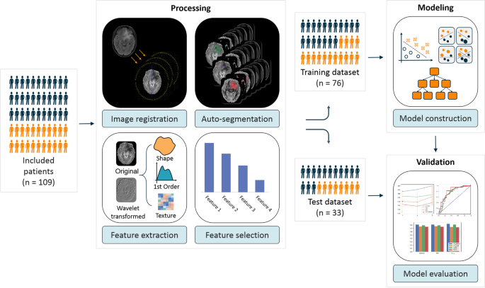
Materials and methods: In this retrospective study, NODDI images were curated from 109 patients with Gb (n = 57) or SBM (n = 52). Automatically segmented multiple volumes of interest (VOIs) encompassed the main tumor regions, including necrosis, solid tumor, and peritumoral edema. Radiomics features were extracted for each main tumor region, using three NODDI parameter maps. Radiomics models were developed based on these three NODDI parameter maps and their amalgamation to differentiate between Gb and SBM. Additionally, radiomics models were constructed based on morphological magnetic resonance imaging (MRI) and diffusion imaging (diffusion-weighted imaging [DWI]; diffusion tensor imaging [DTI]) for performance comparison.
Results: The validation dataset results revealed that the performance of a single NODDI parameter map model was inferior to that of the combined NODDI model. In the necrotic regions, the combined NODDI radiomics model exhibited less than ideal discriminative capabilities (area under the receiver operating characteristic curve [AUC] = 0.701). For peritumoral edema regions, the combined NODDI radiomics model achieved a moderate level of discrimination (AUC = 0.820). Within the solid tumor regions, the combined NODDI radiomics model demonstrated superior performance (AUC = 0.904), surpassing the models of other VOIs. The comparison results demonstrated that the NODDI model was better than the DWI and DTI models, while those of the morphological MRI and NODDI models were similar.
Conclusion: The NODDI radiomics model showed promising performance for preoperative discrimination between Gb and SBM.
Clinical relevance statement: The NODDI radiomics model showed promising performance for preoperative discrimination between Gb and SBM, and radiomics features can be incorporated into the multidimensional phenotypic features that describe tumor heterogeneity.
Key points: • The neurite orientation dispersion and density imaging (NODDI) radiomics model showed promising performance for preoperative discrimination between glioblastoma and solitary brain metastasis. • Compared with other tumor volumes of interest, the NODDI radiomics model based on solid tumor regions performed best in distinguishing the two types of tumors. • The performance of the single-parameter NODDI model was inferior to that of the combined-parameter NODDI model.
期刊介绍:
European Radiology (ER) continuously updates scientific knowledge in radiology by publication of strong original articles and state-of-the-art reviews written by leading radiologists. A well balanced combination of review articles, original papers, short communications from European radiological congresses and information on society matters makes ER an indispensable source for current information in this field.
This is the Journal of the European Society of Radiology, and the official journal of a number of societies.
From 2004-2008 supplements to European Radiology were published under its companion, European Radiology Supplements, ISSN 1613-3749.


 分享
分享
 求助内容:
求助内容: 应助结果提醒方式:
 扫码关注我们
