Eddy D Zandee van Rilland, Se-Young Yoon, Hillary W Garner, Jennifer Ni Mhuircheartaigh, Jim S Wu
{"title":"是否存在宏观的区域内脂肪可排除恶性肿瘤?对613例经组织学证实的恶性骨病变的分析。","authors":"Eddy D Zandee van Rilland, Se-Young Yoon, Hillary W Garner, Jennifer Ni Mhuircheartaigh, Jim S Wu","doi":"10.1007/s00330-024-10687-7","DOIUrl":null,"url":null,"abstract":"<p><strong>Objective: </strong>To determine if macroscopic intralesional fat detected in bone lesions on CT by Hounsfield unit (HU) measurement and on MRI by macroscopic assessment excludes malignancy.</p><p><strong>Materials and methods: </strong>All consecutive CT-guided core needle biopsies (CNB) of non-spinal bone lesions performed at a tertiary center between December 2005 and September 2021 were reviewed. Demographic and histopathology data were recorded. All cases with malignant histopathology were selected, and imaging studies were reviewed. Two independent readers performed CT HU measurements on all bone lesions using a circular region of interest (ROI) to quantitate intralesional fat density (mean HU < -30). MRI images were reviewed to qualitatively assess for macroscopic intralesional fat signal in a subset of patients. Inter-reader agreement was assessed with Cronbach's alpha and intraclass correlation coefficient.</p><p><strong>Results: </strong>In 613 patients (mean age 62.9 years (range 19-95 years), 47.6% female), CT scans from the CNB of 613 malignant bone lesions were reviewed, and 212 cases had additional MRI images. Only 3 cases (0.5%) demonstrated macroscopic intralesional fat on either CT or MRI. One case demonstrated macroscopic intralesional fat density on CT in a case of metastatic prostate cancer. Two cases demonstrated macroscopic intralesional fat signal on MRI in cases of chondrosarcoma and osteosarcoma. Inter-reader agreement was excellent (Cronbach's alpha, 0.95-0.98; intraclass correlation coefficient, 0.90-0.97).</p><p><strong>Conclusion: </strong>Malignant lesions rarely contain macroscopic intralesional fat on CT or MRI. While CT is effective in detecting macroscopic intralesional fat in primarily lytic lesions, MRI may be better for the assessment of heterogenous and infiltrative lesions with mixed lytic and sclerotic components.</p><p><strong>Clinical relevance statement: </strong>Macroscopic intralesional fat is rarely seen in malignant bone tumors and its presence can help to guide the diagnostic workup of bone lesions.</p><p><strong>Key points: </strong>• Presence of macroscopic intralesional fat in bone lesions has been widely theorized as a sign of benignity, but there is limited supporting evidence in the literature. • CT and MRI are effective in evaluating for macroscopic intralesional fat in malignant bone lesions with excellent inter-reader agreement. • Macroscopic intralesional fat is rarely seen in malignant bone lesions.</p>","PeriodicalId":12076,"journal":{"name":"European Radiology","volume":null,"pages":null},"PeriodicalIF":4.7000,"publicationDate":"2024-10-01","publicationTypes":"Journal Article","fieldsOfStudy":null,"isOpenAccess":false,"openAccessPdf":"","citationCount":"0","resultStr":"{\"title\":\"Does the presence of macroscopic intralesional fat exclude malignancy? An analysis of 613 histologically proven malignant bone lesions.\",\"authors\":\"Eddy D Zandee van Rilland, Se-Young Yoon, Hillary W Garner, Jennifer Ni Mhuircheartaigh, Jim S Wu\",\"doi\":\"10.1007/s00330-024-10687-7\",\"DOIUrl\":null,\"url\":null,\"abstract\":\"<p><strong>Objective: </strong>To determine if macroscopic intralesional fat detected in bone lesions on CT by Hounsfield unit (HU) measurement and on MRI by macroscopic assessment excludes malignancy.</p><p><strong>Materials and methods: </strong>All consecutive CT-guided core needle biopsies (CNB) of non-spinal bone lesions performed at a tertiary center between December 2005 and September 2021 were reviewed. Demographic and histopathology data were recorded. All cases with malignant histopathology were selected, and imaging studies were reviewed. Two independent readers performed CT HU measurements on all bone lesions using a circular region of interest (ROI) to quantitate intralesional fat density (mean HU < -30). MRI images were reviewed to qualitatively assess for macroscopic intralesional fat signal in a subset of patients. Inter-reader agreement was assessed with Cronbach's alpha and intraclass correlation coefficient.</p><p><strong>Results: </strong>In 613 patients (mean age 62.9 years (range 19-95 years), 47.6% female), CT scans from the CNB of 613 malignant bone lesions were reviewed, and 212 cases had additional MRI images. Only 3 cases (0.5%) demonstrated macroscopic intralesional fat on either CT or MRI. One case demonstrated macroscopic intralesional fat density on CT in a case of metastatic prostate cancer. Two cases demonstrated macroscopic intralesional fat signal on MRI in cases of chondrosarcoma and osteosarcoma. Inter-reader agreement was excellent (Cronbach's alpha, 0.95-0.98; intraclass correlation coefficient, 0.90-0.97).</p><p><strong>Conclusion: </strong>Malignant lesions rarely contain macroscopic intralesional fat on CT or MRI. While CT is effective in detecting macroscopic intralesional fat in primarily lytic lesions, MRI may be better for the assessment of heterogenous and infiltrative lesions with mixed lytic and sclerotic components.</p><p><strong>Clinical relevance statement: </strong>Macroscopic intralesional fat is rarely seen in malignant bone tumors and its presence can help to guide the diagnostic workup of bone lesions.</p><p><strong>Key points: </strong>• Presence of macroscopic intralesional fat in bone lesions has been widely theorized as a sign of benignity, but there is limited supporting evidence in the literature. • CT and MRI are effective in evaluating for macroscopic intralesional fat in malignant bone lesions with excellent inter-reader agreement. • Macroscopic intralesional fat is rarely seen in malignant bone lesions.</p>\",\"PeriodicalId\":12076,\"journal\":{\"name\":\"European Radiology\",\"volume\":null,\"pages\":null},\"PeriodicalIF\":4.7000,\"publicationDate\":\"2024-10-01\",\"publicationTypes\":\"Journal Article\",\"fieldsOfStudy\":null,\"isOpenAccess\":false,\"openAccessPdf\":\"\",\"citationCount\":\"0\",\"resultStr\":null,\"platform\":\"Semanticscholar\",\"paperid\":null,\"PeriodicalName\":\"European Radiology\",\"FirstCategoryId\":\"3\",\"ListUrlMain\":\"https://doi.org/10.1007/s00330-024-10687-7\",\"RegionNum\":2,\"RegionCategory\":\"医学\",\"ArticlePicture\":[],\"TitleCN\":null,\"AbstractTextCN\":null,\"PMCID\":null,\"EPubDate\":\"2024/3/15 0:00:00\",\"PubModel\":\"Epub\",\"JCR\":\"Q1\",\"JCRName\":\"RADIOLOGY, NUCLEAR MEDICINE & MEDICAL IMAGING\",\"Score\":null,\"Total\":0}","platform":"Semanticscholar","paperid":null,"PeriodicalName":"European Radiology","FirstCategoryId":"3","ListUrlMain":"https://doi.org/10.1007/s00330-024-10687-7","RegionNum":2,"RegionCategory":"医学","ArticlePicture":[],"TitleCN":null,"AbstractTextCN":null,"PMCID":null,"EPubDate":"2024/3/15 0:00:00","PubModel":"Epub","JCR":"Q1","JCRName":"RADIOLOGY, NUCLEAR MEDICINE & MEDICAL IMAGING","Score":null,"Total":0}
Does the presence of macroscopic intralesional fat exclude malignancy? An analysis of 613 histologically proven malignant bone lesions.
Objective: To determine if macroscopic intralesional fat detected in bone lesions on CT by Hounsfield unit (HU) measurement and on MRI by macroscopic assessment excludes malignancy.
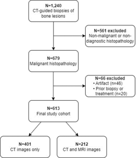
Materials and methods: All consecutive CT-guided core needle biopsies (CNB) of non-spinal bone lesions performed at a tertiary center between December 2005 and September 2021 were reviewed. Demographic and histopathology data were recorded. All cases with malignant histopathology were selected, and imaging studies were reviewed. Two independent readers performed CT HU measurements on all bone lesions using a circular region of interest (ROI) to quantitate intralesional fat density (mean HU < -30). MRI images were reviewed to qualitatively assess for macroscopic intralesional fat signal in a subset of patients. Inter-reader agreement was assessed with Cronbach's alpha and intraclass correlation coefficient.
Results: In 613 patients (mean age 62.9 years (range 19-95 years), 47.6% female), CT scans from the CNB of 613 malignant bone lesions were reviewed, and 212 cases had additional MRI images. Only 3 cases (0.5%) demonstrated macroscopic intralesional fat on either CT or MRI. One case demonstrated macroscopic intralesional fat density on CT in a case of metastatic prostate cancer. Two cases demonstrated macroscopic intralesional fat signal on MRI in cases of chondrosarcoma and osteosarcoma. Inter-reader agreement was excellent (Cronbach's alpha, 0.95-0.98; intraclass correlation coefficient, 0.90-0.97).
Conclusion: Malignant lesions rarely contain macroscopic intralesional fat on CT or MRI. While CT is effective in detecting macroscopic intralesional fat in primarily lytic lesions, MRI may be better for the assessment of heterogenous and infiltrative lesions with mixed lytic and sclerotic components.
Clinical relevance statement: Macroscopic intralesional fat is rarely seen in malignant bone tumors and its presence can help to guide the diagnostic workup of bone lesions.
Key points: • Presence of macroscopic intralesional fat in bone lesions has been widely theorized as a sign of benignity, but there is limited supporting evidence in the literature. • CT and MRI are effective in evaluating for macroscopic intralesional fat in malignant bone lesions with excellent inter-reader agreement. • Macroscopic intralesional fat is rarely seen in malignant bone lesions.
期刊介绍:
European Radiology (ER) continuously updates scientific knowledge in radiology by publication of strong original articles and state-of-the-art reviews written by leading radiologists. A well balanced combination of review articles, original papers, short communications from European radiological congresses and information on society matters makes ER an indispensable source for current information in this field.
This is the Journal of the European Society of Radiology, and the official journal of a number of societies.
From 2004-2008 supplements to European Radiology were published under its companion, European Radiology Supplements, ISSN 1613-3749.


 分享
分享
 求助内容:
求助内容: 应助结果提醒方式:
 扫码关注我们
