Florian Hinterwimmer, Ricardo Smits Serena, Nikolas Wilhelm, Sebastian Breden, Sarah Consalvo, Fritz Seidl, Dominik Juestel, Rainer H H Burgkart, Klaus Woertler, Ruediger von Eisenhart-Rothe, Jan Neumann, Daniel Rueckert
{"title":"基于推荐的骨肿瘤分类与射线照片--与过去的联系。","authors":"Florian Hinterwimmer, Ricardo Smits Serena, Nikolas Wilhelm, Sebastian Breden, Sarah Consalvo, Fritz Seidl, Dominik Juestel, Rainer H H Burgkart, Klaus Woertler, Ruediger von Eisenhart-Rothe, Jan Neumann, Daniel Rueckert","doi":"10.1007/s00330-024-10672-0","DOIUrl":null,"url":null,"abstract":"<p><strong>Objectives: </strong>To develop an algorithm to link undiagnosed patients to previous patient histories based on radiographs, and simultaneous classification of multiple bone tumours to enable early and specific diagnosis.</p><p><strong>Materials and methods: </strong>For this retrospective study, data from 2000 to 2021 were curated from our database by two orthopaedic surgeons, a radiologist and a data scientist. Patients with complete clinical and pre-therapy radiographic data were eligible. To ensure feasibility, the ten most frequent primary tumour entities, confirmed histologically or by tumour board decision, were included. We implemented a ResNet and transformer model to establish baseline results. Our method extracts image features using deep learning and then clusters the k most similar images to the target image using a hash-based nearest-neighbour recommender approach that performs simultaneous classification by majority voting. The results were evaluated with precision-at-k, accuracy, precision and recall. Discrete parameters were described by incidence and percentage ratios. For continuous parameters, based on a normality test, respective statistical measures were calculated.</p><p><strong>Results: </strong>Included were data from 809 patients (1792 radiographs; mean age 33.73 ± 18.65, range 3-89 years; 443 men), with Osteochondroma (28.31%) and Ewing sarcoma (1.11%) as the most and least common entities, respectively. The dataset was split into training (80%) and test subsets (20%). For k = 3, our model achieved the highest mean accuracy, precision and recall (92.86%, 92.86% and 34.08%), significantly outperforming state-of-the-art models (54.10%, 55.57%, 19.85% and 62.80%, 61.33%, 23.05%).</p><p><strong>Conclusion: </strong>Our novel approach surpasses current models in tumour classification and links to past patient data, leveraging expert insights.</p><p><strong>Clinical relevance statement: </strong>The proposed algorithm could serve as a vital support tool for clinicians and general practitioners with limited experience in bone tumour classification by identifying similar cases and classifying bone tumour entities.</p><p><strong>Key points: </strong>• Addressed accurate bone tumour classification using radiographic features. • Model achieved 92.86%, 92.86% and 34.08% mean accuracy, precision and recall, respectively, significantly surpassing state-of-the-art models. • Enhanced diagnosis by integrating prior expert patient assessments.</p>","PeriodicalId":12076,"journal":{"name":"European Radiology","volume":" ","pages":"6629-6638"},"PeriodicalIF":4.7000,"publicationDate":"2024-10-01","publicationTypes":"Journal Article","fieldsOfStudy":null,"isOpenAccess":false,"openAccessPdf":"https://www.ncbi.nlm.nih.gov/pmc/articles/PMC11399296/pdf/","citationCount":"0","resultStr":"{\"title\":\"Recommender-based bone tumour classification with radiographs-a link to the past.\",\"authors\":\"Florian Hinterwimmer, Ricardo Smits Serena, Nikolas Wilhelm, Sebastian Breden, Sarah Consalvo, Fritz Seidl, Dominik Juestel, Rainer H H Burgkart, Klaus Woertler, Ruediger von Eisenhart-Rothe, Jan Neumann, Daniel Rueckert\",\"doi\":\"10.1007/s00330-024-10672-0\",\"DOIUrl\":null,\"url\":null,\"abstract\":\"<p><strong>Objectives: </strong>To develop an algorithm to link undiagnosed patients to previous patient histories based on radiographs, and simultaneous classification of multiple bone tumours to enable early and specific diagnosis.</p><p><strong>Materials and methods: </strong>For this retrospective study, data from 2000 to 2021 were curated from our database by two orthopaedic surgeons, a radiologist and a data scientist. Patients with complete clinical and pre-therapy radiographic data were eligible. To ensure feasibility, the ten most frequent primary tumour entities, confirmed histologically or by tumour board decision, were included. We implemented a ResNet and transformer model to establish baseline results. Our method extracts image features using deep learning and then clusters the k most similar images to the target image using a hash-based nearest-neighbour recommender approach that performs simultaneous classification by majority voting. The results were evaluated with precision-at-k, accuracy, precision and recall. Discrete parameters were described by incidence and percentage ratios. For continuous parameters, based on a normality test, respective statistical measures were calculated.</p><p><strong>Results: </strong>Included were data from 809 patients (1792 radiographs; mean age 33.73 ± 18.65, range 3-89 years; 443 men), with Osteochondroma (28.31%) and Ewing sarcoma (1.11%) as the most and least common entities, respectively. The dataset was split into training (80%) and test subsets (20%). For k = 3, our model achieved the highest mean accuracy, precision and recall (92.86%, 92.86% and 34.08%), significantly outperforming state-of-the-art models (54.10%, 55.57%, 19.85% and 62.80%, 61.33%, 23.05%).</p><p><strong>Conclusion: </strong>Our novel approach surpasses current models in tumour classification and links to past patient data, leveraging expert insights.</p><p><strong>Clinical relevance statement: </strong>The proposed algorithm could serve as a vital support tool for clinicians and general practitioners with limited experience in bone tumour classification by identifying similar cases and classifying bone tumour entities.</p><p><strong>Key points: </strong>• Addressed accurate bone tumour classification using radiographic features. • Model achieved 92.86%, 92.86% and 34.08% mean accuracy, precision and recall, respectively, significantly surpassing state-of-the-art models. • Enhanced diagnosis by integrating prior expert patient assessments.</p>\",\"PeriodicalId\":12076,\"journal\":{\"name\":\"European Radiology\",\"volume\":\" \",\"pages\":\"6629-6638\"},\"PeriodicalIF\":4.7000,\"publicationDate\":\"2024-10-01\",\"publicationTypes\":\"Journal Article\",\"fieldsOfStudy\":null,\"isOpenAccess\":false,\"openAccessPdf\":\"https://www.ncbi.nlm.nih.gov/pmc/articles/PMC11399296/pdf/\",\"citationCount\":\"0\",\"resultStr\":null,\"platform\":\"Semanticscholar\",\"paperid\":null,\"PeriodicalName\":\"European Radiology\",\"FirstCategoryId\":\"3\",\"ListUrlMain\":\"https://doi.org/10.1007/s00330-024-10672-0\",\"RegionNum\":2,\"RegionCategory\":\"医学\",\"ArticlePicture\":[],\"TitleCN\":null,\"AbstractTextCN\":null,\"PMCID\":null,\"EPubDate\":\"2024/3/15 0:00:00\",\"PubModel\":\"Epub\",\"JCR\":\"Q1\",\"JCRName\":\"RADIOLOGY, NUCLEAR MEDICINE & MEDICAL IMAGING\",\"Score\":null,\"Total\":0}","platform":"Semanticscholar","paperid":null,"PeriodicalName":"European Radiology","FirstCategoryId":"3","ListUrlMain":"https://doi.org/10.1007/s00330-024-10672-0","RegionNum":2,"RegionCategory":"医学","ArticlePicture":[],"TitleCN":null,"AbstractTextCN":null,"PMCID":null,"EPubDate":"2024/3/15 0:00:00","PubModel":"Epub","JCR":"Q1","JCRName":"RADIOLOGY, NUCLEAR MEDICINE & MEDICAL IMAGING","Score":null,"Total":0}
Recommender-based bone tumour classification with radiographs-a link to the past.
Objectives: To develop an algorithm to link undiagnosed patients to previous patient histories based on radiographs, and simultaneous classification of multiple bone tumours to enable early and specific diagnosis.
Materials and methods: For this retrospective study, data from 2000 to 2021 were curated from our database by two orthopaedic surgeons, a radiologist and a data scientist. Patients with complete clinical and pre-therapy radiographic data were eligible. To ensure feasibility, the ten most frequent primary tumour entities, confirmed histologically or by tumour board decision, were included. We implemented a ResNet and transformer model to establish baseline results. Our method extracts image features using deep learning and then clusters the k most similar images to the target image using a hash-based nearest-neighbour recommender approach that performs simultaneous classification by majority voting. The results were evaluated with precision-at-k, accuracy, precision and recall. Discrete parameters were described by incidence and percentage ratios. For continuous parameters, based on a normality test, respective statistical measures were calculated.
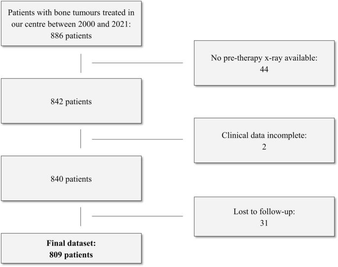
Results: Included were data from 809 patients (1792 radiographs; mean age 33.73 ± 18.65, range 3-89 years; 443 men), with Osteochondroma (28.31%) and Ewing sarcoma (1.11%) as the most and least common entities, respectively. The dataset was split into training (80%) and test subsets (20%). For k = 3, our model achieved the highest mean accuracy, precision and recall (92.86%, 92.86% and 34.08%), significantly outperforming state-of-the-art models (54.10%, 55.57%, 19.85% and 62.80%, 61.33%, 23.05%).
Conclusion: Our novel approach surpasses current models in tumour classification and links to past patient data, leveraging expert insights.
Clinical relevance statement: The proposed algorithm could serve as a vital support tool for clinicians and general practitioners with limited experience in bone tumour classification by identifying similar cases and classifying bone tumour entities.
Key points: • Addressed accurate bone tumour classification using radiographic features. • Model achieved 92.86%, 92.86% and 34.08% mean accuracy, precision and recall, respectively, significantly surpassing state-of-the-art models. • Enhanced diagnosis by integrating prior expert patient assessments.
期刊介绍:
European Radiology (ER) continuously updates scientific knowledge in radiology by publication of strong original articles and state-of-the-art reviews written by leading radiologists. A well balanced combination of review articles, original papers, short communications from European radiological congresses and information on society matters makes ER an indispensable source for current information in this field.
This is the Journal of the European Society of Radiology, and the official journal of a number of societies.
From 2004-2008 supplements to European Radiology were published under its companion, European Radiology Supplements, ISSN 1613-3749.


 分享
分享
 求助内容:
求助内容: 应助结果提醒方式:
 扫码关注我们
