{"title":"皮肤淋巴组织增生性疾病:回到未来。","authors":"Rein Willemze MD","doi":"10.1111/cup.14609","DOIUrl":null,"url":null,"abstract":"<p>In the 1980s, immunohistochemistry and clonality analyses became instrumental in the recognition and definition of new types of cutaneous T-cell lymphoma (CTCL) and cutaneous B-cell lymphoma (CBCL) and the development of new classifications. By accepting loss of pan-T-cell antigens and clonal T-cell receptor gene rearrangements as important criteria to differentiate between benign and malignant T-cell proliferations, and monotypic immunoglobulin light-chain expression and clonal immunoglobulin gene rearrangements as crucial criteria to distinguish between benign and malignant B-cell proliferations, many cases, until then diagnosed as cutaneous lymphoid hyperplasia or pseudolymphoma, were reclassified as primary cutaneous CD4+ small/medium T-cell lymphoma (PCSM-TCL) or primary cutaneous marginal zone lymphoma (PCMZL), respectively. However, in recent years there is growing awareness that neither these immunohistochemical criteria nor demonstration of T-cell or B-cell clonality is specific for malignant lymphomas. In addition, many studies have reported that these low-grade malignant CTCL and CBCL have an indolent clinical behavior and an excellent prognosis with disease-specific survival rates of or close to 100%. As a result, recent classifications have downgraded several low-grade malignant cutaneous lymphomas to lymphoproliferative disorder (LPD). Both the 5th edition of the WHO classification (2022) and the 2022 International Consensus Classification (ICC) of mature lymphoid neoplasms reclassified PCSM-TCL as primary cutaneous CD4+ small/medium T-cell LPD and primary cutaneous acral CD8+ T-cell lymphoma as primary cutaneous acral CD8+ T cell LPD. While the 2022 ICC introduced the term “primary cutaneous marginal zone LPD,” in the 5th edition of the WHO classification PCMZL is maintained. In this review we describe the background and rationale of the continually changing terminology of these conditions and discuss the clinical consequences of downgrading malignant lymphomas to LPDs.</p>","PeriodicalId":15407,"journal":{"name":"Journal of Cutaneous Pathology","volume":"51 6","pages":"468-476"},"PeriodicalIF":1.1000,"publicationDate":"2024-03-18","publicationTypes":"Journal Article","fieldsOfStudy":null,"isOpenAccess":false,"openAccessPdf":"https://onlinelibrary.wiley.com/doi/epdf/10.1111/cup.14609","citationCount":"0","resultStr":"{\"title\":\"Cutaneous lymphoproliferative disorders: Back to the future\",\"authors\":\"Rein Willemze MD\",\"doi\":\"10.1111/cup.14609\",\"DOIUrl\":null,\"url\":null,\"abstract\":\"<p>In the 1980s, immunohistochemistry and clonality analyses became instrumental in the recognition and definition of new types of cutaneous T-cell lymphoma (CTCL) and cutaneous B-cell lymphoma (CBCL) and the development of new classifications. By accepting loss of pan-T-cell antigens and clonal T-cell receptor gene rearrangements as important criteria to differentiate between benign and malignant T-cell proliferations, and monotypic immunoglobulin light-chain expression and clonal immunoglobulin gene rearrangements as crucial criteria to distinguish between benign and malignant B-cell proliferations, many cases, until then diagnosed as cutaneous lymphoid hyperplasia or pseudolymphoma, were reclassified as primary cutaneous CD4+ small/medium T-cell lymphoma (PCSM-TCL) or primary cutaneous marginal zone lymphoma (PCMZL), respectively. However, in recent years there is growing awareness that neither these immunohistochemical criteria nor demonstration of T-cell or B-cell clonality is specific for malignant lymphomas. In addition, many studies have reported that these low-grade malignant CTCL and CBCL have an indolent clinical behavior and an excellent prognosis with disease-specific survival rates of or close to 100%. As a result, recent classifications have downgraded several low-grade malignant cutaneous lymphomas to lymphoproliferative disorder (LPD). Both the 5th edition of the WHO classification (2022) and the 2022 International Consensus Classification (ICC) of mature lymphoid neoplasms reclassified PCSM-TCL as primary cutaneous CD4+ small/medium T-cell LPD and primary cutaneous acral CD8+ T-cell lymphoma as primary cutaneous acral CD8+ T cell LPD. While the 2022 ICC introduced the term “primary cutaneous marginal zone LPD,” in the 5th edition of the WHO classification PCMZL is maintained. In this review we describe the background and rationale of the continually changing terminology of these conditions and discuss the clinical consequences of downgrading malignant lymphomas to LPDs.</p>\",\"PeriodicalId\":15407,\"journal\":{\"name\":\"Journal of Cutaneous Pathology\",\"volume\":\"51 6\",\"pages\":\"468-476\"},\"PeriodicalIF\":1.1000,\"publicationDate\":\"2024-03-18\",\"publicationTypes\":\"Journal Article\",\"fieldsOfStudy\":null,\"isOpenAccess\":false,\"openAccessPdf\":\"https://onlinelibrary.wiley.com/doi/epdf/10.1111/cup.14609\",\"citationCount\":\"0\",\"resultStr\":null,\"platform\":\"Semanticscholar\",\"paperid\":null,\"PeriodicalName\":\"Journal of Cutaneous Pathology\",\"FirstCategoryId\":\"3\",\"ListUrlMain\":\"https://onlinelibrary.wiley.com/doi/10.1111/cup.14609\",\"RegionNum\":4,\"RegionCategory\":\"医学\",\"ArticlePicture\":[],\"TitleCN\":null,\"AbstractTextCN\":null,\"PMCID\":null,\"EPubDate\":\"\",\"PubModel\":\"\",\"JCR\":\"Q3\",\"JCRName\":\"DERMATOLOGY\",\"Score\":null,\"Total\":0}","platform":"Semanticscholar","paperid":null,"PeriodicalName":"Journal of Cutaneous Pathology","FirstCategoryId":"3","ListUrlMain":"https://onlinelibrary.wiley.com/doi/10.1111/cup.14609","RegionNum":4,"RegionCategory":"医学","ArticlePicture":[],"TitleCN":null,"AbstractTextCN":null,"PMCID":null,"EPubDate":"","PubModel":"","JCR":"Q3","JCRName":"DERMATOLOGY","Score":null,"Total":0}
引用次数: 0
摘要
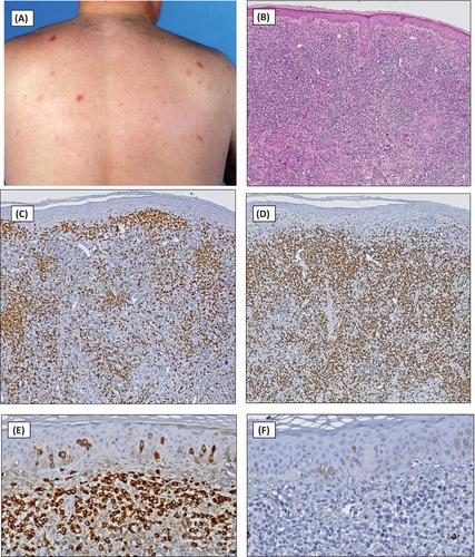
20 世纪 80 年代,免疫组化和克隆性分析在识别和定义新类型的皮肤 T 细胞淋巴瘤(CTCL)和皮肤 B 细胞淋巴瘤(CBCL)以及制定新的分类标准方面发挥了重要作用。通过接受泛T细胞抗原缺失和克隆T细胞受体基因重排作为区分良性和恶性T细胞增生的重要标准,以及接受单型免疫球蛋白轻链表达和克隆免疫球蛋白基因重排作为区分良性和恶性B细胞增生的重要标准,许多病例在此之前被诊断为皮肤T细胞淋巴瘤(CTCL)和皮肤B细胞淋巴瘤(CBCL)、在此之前,许多被诊断为皮肤淋巴样增生或假淋巴瘤的病例被重新分类为原发性皮肤 CD4+ 小/中 T 细胞淋巴瘤(PCSM-TCL)或原发性皮肤边缘区淋巴瘤(PCMZL)。然而,近年来人们越来越认识到,无论是这些免疫组化标准,还是 T 细胞或 B 细胞克隆性的证明,都不是恶性淋巴瘤的特异性标准。此外,许多研究报告称,这些低度恶性 CTCL 和 CBCL 临床表现不明显,预后良好,疾病特异性生存率达到或接近 100%。因此,最近的分类将几种低度恶性皮肤淋巴瘤降级为淋巴增生性疾病(LPD)。第五版世卫组织分类(2022 年)和 2022 年成熟淋巴肿瘤国际共识分类(ICC)都将 PCSM-TCL 重新分类为原发性皮肤 CD4+ 小/中 T 细胞淋巴增生性疾病,将原发性皮肤尖锐湿疣 CD8+ T 细胞淋巴瘤重新分类为原发性皮肤尖锐湿疣 CD8+ T 细胞淋巴增生性疾病。2022 年 ICC 引入了 "原发性皮肤边缘区 LPD "这一术语,而在第五版世界卫生组织分类中则保留了 PCMZL。在这篇综述中,我们描述了这些疾病术语不断变化的背景和原因,并讨论了将恶性淋巴瘤降级为 LPD 的临床后果。
Cutaneous lymphoproliferative disorders: Back to the future
In the 1980s, immunohistochemistry and clonality analyses became instrumental in the recognition and definition of new types of cutaneous T-cell lymphoma (CTCL) and cutaneous B-cell lymphoma (CBCL) and the development of new classifications. By accepting loss of pan-T-cell antigens and clonal T-cell receptor gene rearrangements as important criteria to differentiate between benign and malignant T-cell proliferations, and monotypic immunoglobulin light-chain expression and clonal immunoglobulin gene rearrangements as crucial criteria to distinguish between benign and malignant B-cell proliferations, many cases, until then diagnosed as cutaneous lymphoid hyperplasia or pseudolymphoma, were reclassified as primary cutaneous CD4+ small/medium T-cell lymphoma (PCSM-TCL) or primary cutaneous marginal zone lymphoma (PCMZL), respectively. However, in recent years there is growing awareness that neither these immunohistochemical criteria nor demonstration of T-cell or B-cell clonality is specific for malignant lymphomas. In addition, many studies have reported that these low-grade malignant CTCL and CBCL have an indolent clinical behavior and an excellent prognosis with disease-specific survival rates of or close to 100%. As a result, recent classifications have downgraded several low-grade malignant cutaneous lymphomas to lymphoproliferative disorder (LPD). Both the 5th edition of the WHO classification (2022) and the 2022 International Consensus Classification (ICC) of mature lymphoid neoplasms reclassified PCSM-TCL as primary cutaneous CD4+ small/medium T-cell LPD and primary cutaneous acral CD8+ T-cell lymphoma as primary cutaneous acral CD8+ T cell LPD. While the 2022 ICC introduced the term “primary cutaneous marginal zone LPD,” in the 5th edition of the WHO classification PCMZL is maintained. In this review we describe the background and rationale of the continually changing terminology of these conditions and discuss the clinical consequences of downgrading malignant lymphomas to LPDs.
期刊介绍:
Journal of Cutaneous Pathology publishes manuscripts broadly relevant to diseases of the skin and mucosae, with the aims of advancing scientific knowledge regarding dermatopathology and enhancing the communication between clinical practitioners and research scientists. Original scientific manuscripts on diagnostic and experimental cutaneous pathology are especially desirable. Timely, pertinent review articles also will be given high priority. Manuscripts based on light, fluorescence, and electron microscopy, histochemistry, immunology, molecular biology, and genetics, as well as allied sciences, are all welcome, provided their principal focus is on cutaneous pathology. Publication time will be kept as short as possible, ensuring that articles will be quickly available to all interested in this speciality.


 分享
分享
 求助内容:
求助内容: 应助结果提醒方式:
 扫码关注我们
