Christopher S Morrow, Kelsey Tweed, Sabina Farhadova, Alex J Walsh, Bo P Lear, Avtar Roopra, Ryan D Risgaard, Payton C Klosa, Zachary P Arndt, Ella R Peterson, Michelle M Chi, Allison G Harris, Melissa C Skala, Darcie L Moore
{"title":"自发荧光是神经干细胞活化状态的生物标记。","authors":"Christopher S Morrow, Kelsey Tweed, Sabina Farhadova, Alex J Walsh, Bo P Lear, Avtar Roopra, Ryan D Risgaard, Payton C Klosa, Zachary P Arndt, Ella R Peterson, Michelle M Chi, Allison G Harris, Melissa C Skala, Darcie L Moore","doi":"10.1016/j.stem.2024.02.011","DOIUrl":null,"url":null,"abstract":"<p><p>Neural stem cells (NSCs) must exit quiescence to produce neurons; however, our understanding of this process remains constrained by the technical limitations of current technologies. Fluorescence lifetime imaging (FLIM) of autofluorescent metabolic cofactors has been used in other cell types to study shifts in cell states driven by metabolic remodeling that change the optical properties of these endogenous fluorophores. Using this non-destructive, live-cell, and label-free strategy, we found that quiescent NSCs (qNSCs) and activated NSCs (aNSCs) have unique autofluorescence profiles. Specifically, qNSCs display an enrichment of autofluorescence localizing to a subset of lysosomes, which can be used as a graded marker of NSC quiescence to predict cell behavior at single-cell resolution. Coupling autofluorescence imaging with single-cell RNA sequencing, we provide resources revealing transcriptional features linked to deep quiescence and rapid NSC activation. Together, we describe an approach for tracking mouse NSC activation state and expand our understanding of adult neurogenesis.</p>","PeriodicalId":93928,"journal":{"name":"Cell stem cell","volume":" ","pages":"570-581.e7"},"PeriodicalIF":20.4000,"publicationDate":"2024-04-04","publicationTypes":"Journal Article","fieldsOfStudy":null,"isOpenAccess":false,"openAccessPdf":"https://www.ncbi.nlm.nih.gov/pmc/articles/PMC10997463/pdf/","citationCount":"0","resultStr":"{\"title\":\"Autofluorescence is a biomarker of neural stem cell activation state.\",\"authors\":\"Christopher S Morrow, Kelsey Tweed, Sabina Farhadova, Alex J Walsh, Bo P Lear, Avtar Roopra, Ryan D Risgaard, Payton C Klosa, Zachary P Arndt, Ella R Peterson, Michelle M Chi, Allison G Harris, Melissa C Skala, Darcie L Moore\",\"doi\":\"10.1016/j.stem.2024.02.011\",\"DOIUrl\":null,\"url\":null,\"abstract\":\"<p><p>Neural stem cells (NSCs) must exit quiescence to produce neurons; however, our understanding of this process remains constrained by the technical limitations of current technologies. Fluorescence lifetime imaging (FLIM) of autofluorescent metabolic cofactors has been used in other cell types to study shifts in cell states driven by metabolic remodeling that change the optical properties of these endogenous fluorophores. Using this non-destructive, live-cell, and label-free strategy, we found that quiescent NSCs (qNSCs) and activated NSCs (aNSCs) have unique autofluorescence profiles. Specifically, qNSCs display an enrichment of autofluorescence localizing to a subset of lysosomes, which can be used as a graded marker of NSC quiescence to predict cell behavior at single-cell resolution. Coupling autofluorescence imaging with single-cell RNA sequencing, we provide resources revealing transcriptional features linked to deep quiescence and rapid NSC activation. Together, we describe an approach for tracking mouse NSC activation state and expand our understanding of adult neurogenesis.</p>\",\"PeriodicalId\":93928,\"journal\":{\"name\":\"Cell stem cell\",\"volume\":\" \",\"pages\":\"570-581.e7\"},\"PeriodicalIF\":20.4000,\"publicationDate\":\"2024-04-04\",\"publicationTypes\":\"Journal Article\",\"fieldsOfStudy\":null,\"isOpenAccess\":false,\"openAccessPdf\":\"https://www.ncbi.nlm.nih.gov/pmc/articles/PMC10997463/pdf/\",\"citationCount\":\"0\",\"resultStr\":null,\"platform\":\"Semanticscholar\",\"paperid\":null,\"PeriodicalName\":\"Cell stem cell\",\"FirstCategoryId\":\"1085\",\"ListUrlMain\":\"https://doi.org/10.1016/j.stem.2024.02.011\",\"RegionNum\":0,\"RegionCategory\":null,\"ArticlePicture\":[],\"TitleCN\":null,\"AbstractTextCN\":null,\"PMCID\":null,\"EPubDate\":\"2024/3/22 0:00:00\",\"PubModel\":\"Epub\",\"JCR\":\"\",\"JCRName\":\"\",\"Score\":null,\"Total\":0}","platform":"Semanticscholar","paperid":null,"PeriodicalName":"Cell stem cell","FirstCategoryId":"1085","ListUrlMain":"https://doi.org/10.1016/j.stem.2024.02.011","RegionNum":0,"RegionCategory":null,"ArticlePicture":[],"TitleCN":null,"AbstractTextCN":null,"PMCID":null,"EPubDate":"2024/3/22 0:00:00","PubModel":"Epub","JCR":"","JCRName":"","Score":null,"Total":0}
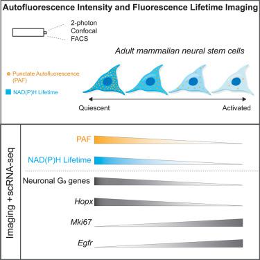
Autofluorescence is a biomarker of neural stem cell activation state.
Neural stem cells (NSCs) must exit quiescence to produce neurons; however, our understanding of this process remains constrained by the technical limitations of current technologies. Fluorescence lifetime imaging (FLIM) of autofluorescent metabolic cofactors has been used in other cell types to study shifts in cell states driven by metabolic remodeling that change the optical properties of these endogenous fluorophores. Using this non-destructive, live-cell, and label-free strategy, we found that quiescent NSCs (qNSCs) and activated NSCs (aNSCs) have unique autofluorescence profiles. Specifically, qNSCs display an enrichment of autofluorescence localizing to a subset of lysosomes, which can be used as a graded marker of NSC quiescence to predict cell behavior at single-cell resolution. Coupling autofluorescence imaging with single-cell RNA sequencing, we provide resources revealing transcriptional features linked to deep quiescence and rapid NSC activation. Together, we describe an approach for tracking mouse NSC activation state and expand our understanding of adult neurogenesis.


 分享
分享

 求助内容:
求助内容: 应助结果提醒方式:
 扫码关注我们
