Yao Zheng, Jingliang Zhang, Dong Huang, Xiaoshuo Hao, Weijun Qin, Yang Liu
{"title":"利用弱监督深度学习模型检测核磁共振成像看不见的前列腺癌","authors":"Yao Zheng, Jingliang Zhang, Dong Huang, Xiaoshuo Hao, Weijun Qin, Yang Liu","doi":"10.1155/2024/2741986","DOIUrl":null,"url":null,"abstract":"<p><strong>Background: </strong>MRI is an important tool for accurate detection and targeted biopsy of prostate lesions. However, the imaging appearances of some prostate cancers are similar to those of the surrounding normal tissue on MRI, which are referred to as MRI-invisible prostate cancers (MIPCas). The detection of MIPCas remains challenging and requires extensive systematic biopsy for identification. In this study, we developed a weakly supervised UNet (WSUNet) to detect MIPCas.</p><p><strong>Methods: </strong>The study included 777 patients (training set: 600; testing set: 177), all of them underwent comprehensive prostate biopsies using an MRI-ultrasound fusion system. MIPCas were identified in MRI based on the Gleason grade (≥7) from known systematic biopsy results.</p><p><strong>Results: </strong>The WSUNet model underwent validation through systematic biopsy in the testing set with an AUC of 0.764 (95% CI: 0.728-0.798). Furthermore, WSUNet exhibited a statistically significant precision improvement of 91.3% (<i>p</i> < 0.01) over conventional systematic biopsy methods in the testing set. This improvement resulted in a substantial 47.6% (<i>p</i> < 0.01) decrease in unnecessary biopsy needles, while maintaining the same number of positively identified cores as in the original systematic biopsy.</p><p><strong>Conclusions: </strong>In conclusion, the proposed WSUNet could effectively detect MIPCas, thereby reducing unnecessary biopsies.</p>","PeriodicalId":47063,"journal":{"name":"International Journal of Biomedical Imaging","volume":"2024 ","pages":"2741986"},"PeriodicalIF":1.3000,"publicationDate":"2024-03-19","publicationTypes":"Journal Article","fieldsOfStudy":null,"isOpenAccess":false,"openAccessPdf":"https://www.ncbi.nlm.nih.gov/pmc/articles/PMC10965281/pdf/","citationCount":"0","resultStr":"{\"title\":\"Detecting MRI-Invisible Prostate Cancers Using a Weakly Supervised Deep Learning Model.\",\"authors\":\"Yao Zheng, Jingliang Zhang, Dong Huang, Xiaoshuo Hao, Weijun Qin, Yang Liu\",\"doi\":\"10.1155/2024/2741986\",\"DOIUrl\":null,\"url\":null,\"abstract\":\"<p><strong>Background: </strong>MRI is an important tool for accurate detection and targeted biopsy of prostate lesions. However, the imaging appearances of some prostate cancers are similar to those of the surrounding normal tissue on MRI, which are referred to as MRI-invisible prostate cancers (MIPCas). The detection of MIPCas remains challenging and requires extensive systematic biopsy for identification. In this study, we developed a weakly supervised UNet (WSUNet) to detect MIPCas.</p><p><strong>Methods: </strong>The study included 777 patients (training set: 600; testing set: 177), all of them underwent comprehensive prostate biopsies using an MRI-ultrasound fusion system. MIPCas were identified in MRI based on the Gleason grade (≥7) from known systematic biopsy results.</p><p><strong>Results: </strong>The WSUNet model underwent validation through systematic biopsy in the testing set with an AUC of 0.764 (95% CI: 0.728-0.798). Furthermore, WSUNet exhibited a statistically significant precision improvement of 91.3% (<i>p</i> < 0.01) over conventional systematic biopsy methods in the testing set. This improvement resulted in a substantial 47.6% (<i>p</i> < 0.01) decrease in unnecessary biopsy needles, while maintaining the same number of positively identified cores as in the original systematic biopsy.</p><p><strong>Conclusions: </strong>In conclusion, the proposed WSUNet could effectively detect MIPCas, thereby reducing unnecessary biopsies.</p>\",\"PeriodicalId\":47063,\"journal\":{\"name\":\"International Journal of Biomedical Imaging\",\"volume\":\"2024 \",\"pages\":\"2741986\"},\"PeriodicalIF\":1.3000,\"publicationDate\":\"2024-03-19\",\"publicationTypes\":\"Journal Article\",\"fieldsOfStudy\":null,\"isOpenAccess\":false,\"openAccessPdf\":\"https://www.ncbi.nlm.nih.gov/pmc/articles/PMC10965281/pdf/\",\"citationCount\":\"0\",\"resultStr\":null,\"platform\":\"Semanticscholar\",\"paperid\":null,\"PeriodicalName\":\"International Journal of Biomedical Imaging\",\"FirstCategoryId\":\"1085\",\"ListUrlMain\":\"https://doi.org/10.1155/2024/2741986\",\"RegionNum\":0,\"RegionCategory\":null,\"ArticlePicture\":[],\"TitleCN\":null,\"AbstractTextCN\":null,\"PMCID\":null,\"EPubDate\":\"2024/1/1 0:00:00\",\"PubModel\":\"eCollection\",\"JCR\":\"Q2\",\"JCRName\":\"ENGINEERING, BIOMEDICAL\",\"Score\":null,\"Total\":0}","platform":"Semanticscholar","paperid":null,"PeriodicalName":"International Journal of Biomedical Imaging","FirstCategoryId":"1085","ListUrlMain":"https://doi.org/10.1155/2024/2741986","RegionNum":0,"RegionCategory":null,"ArticlePicture":[],"TitleCN":null,"AbstractTextCN":null,"PMCID":null,"EPubDate":"2024/1/1 0:00:00","PubModel":"eCollection","JCR":"Q2","JCRName":"ENGINEERING, BIOMEDICAL","Score":null,"Total":0}
Detecting MRI-Invisible Prostate Cancers Using a Weakly Supervised Deep Learning Model.
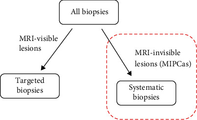
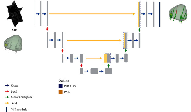
Background: MRI is an important tool for accurate detection and targeted biopsy of prostate lesions. However, the imaging appearances of some prostate cancers are similar to those of the surrounding normal tissue on MRI, which are referred to as MRI-invisible prostate cancers (MIPCas). The detection of MIPCas remains challenging and requires extensive systematic biopsy for identification. In this study, we developed a weakly supervised UNet (WSUNet) to detect MIPCas.
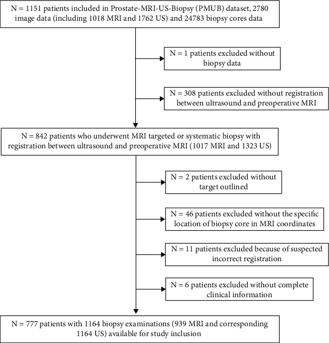
Methods: The study included 777 patients (training set: 600; testing set: 177), all of them underwent comprehensive prostate biopsies using an MRI-ultrasound fusion system. MIPCas were identified in MRI based on the Gleason grade (≥7) from known systematic biopsy results.
Results: The WSUNet model underwent validation through systematic biopsy in the testing set with an AUC of 0.764 (95% CI: 0.728-0.798). Furthermore, WSUNet exhibited a statistically significant precision improvement of 91.3% (p < 0.01) over conventional systematic biopsy methods in the testing set. This improvement resulted in a substantial 47.6% (p < 0.01) decrease in unnecessary biopsy needles, while maintaining the same number of positively identified cores as in the original systematic biopsy.
Conclusions: In conclusion, the proposed WSUNet could effectively detect MIPCas, thereby reducing unnecessary biopsies.
期刊介绍:
The International Journal of Biomedical Imaging is managed by a board of editors comprising internationally renowned active researchers. The journal is freely accessible online and also offered for purchase in print format. It employs a web-based review system to ensure swift turnaround times while maintaining high standards. In addition to regular issues, special issues are organized by guest editors. The subject areas covered include (but are not limited to):
Digital radiography and tomosynthesis
X-ray computed tomography (CT)
Magnetic resonance imaging (MRI)
Single photon emission computed tomography (SPECT)
Positron emission tomography (PET)
Ultrasound imaging
Diffuse optical tomography, coherence, fluorescence, bioluminescence tomography, impedance tomography
Neutron imaging for biomedical applications
Magnetic and optical spectroscopy, and optical biopsy
Optical, electron, scanning tunneling/atomic force microscopy
Small animal imaging
Functional, cellular, and molecular imaging
Imaging assays for screening and molecular analysis
Microarray image analysis and bioinformatics
Emerging biomedical imaging techniques
Imaging modality fusion
Biomedical imaging instrumentation
Biomedical image processing, pattern recognition, and analysis
Biomedical image visualization, compression, transmission, and storage
Imaging and modeling related to systems biology and systems biomedicine
Applied mathematics, applied physics, and chemistry related to biomedical imaging
Grid-enabling technology for biomedical imaging and informatics


 分享
分享
 求助内容:
求助内容: 应助结果提醒方式:
 扫码关注我们
