{"title":"自发回声对比和脐静脉血流减少可预测胎儿腹腔内脐静脉曲张血栓的形成。","authors":"Yu Takaishi, Kaoru Kawasaki, Kazuhiko Uematsu, Shinya Yoshioka","doi":"10.1007/s10396-024-01428-w","DOIUrl":null,"url":null,"abstract":"<p><strong>Purpose: </strong>Fetal intra-abdominal umbilical vein varix (FIUVV) can cause thrombosis, fetal growth restriction (FGR), and intrauterine fetal death (IUFD). However, its management and evaluation to avoid fetal risks have not been elucidated. The aim of this study was to develop a novel method to evaluate fetal risks, including FGR and fetal dysfunction via frequent ultrasound examinations.</p><p><strong>Methods: </strong>A 28-year-old pregnant woman was diagnosed with FIUVV via ultrasound at 26 weeks of gestation and admitted to our hospital. Ultrasound examinations were performed two to three times weekly to evaluate size and shape of the FIUVV and umbilical vein blood flow at the inflow and outflow sites of the FIUVV.</p><p><strong>Results: </strong>The outflow site of the FIUVV was constricted and collapsed, and the blood flow velocity at the inflow site of the FIUVV was decreased. At 32 weeks of gestation, spontaneous echo contrast (SEC), which indicates increased echogenicity, appeared. At 35 weeks of gestation, the patient noticed decreased fetal movement, and CTG showed non-reassuring fetal status. SEC in the FIUVV was remarkable. Fetal movement could not be confirmed at ultrasound. Cesarean section was performed and a 1,854-g healthy infant was delivered with an umbilical cord arterial pH of 7.266.</p><p><strong>Conclusion: </strong>The echographic changes, such as decreased umbilical vein blood flow and SEC, in FIUVV observed in this case could indicate thrombus formation, which can lead to fetal dysfunction. Frequent ultrasound examinations can help determine the timing of delivery and improve the neonatal prognosis.</p>","PeriodicalId":50130,"journal":{"name":"Journal of Medical Ultrasonics","volume":" ","pages":"477-481"},"PeriodicalIF":2.1000,"publicationDate":"2024-07-01","publicationTypes":"Journal Article","fieldsOfStudy":null,"isOpenAccess":false,"openAccessPdf":"","citationCount":"0","resultStr":"{\"title\":\"Spontaneous echo contrast and decreased umbilical vein blood flow may predict thrombus formation in fetal intra-abdominal umbilical vein varix.\",\"authors\":\"Yu Takaishi, Kaoru Kawasaki, Kazuhiko Uematsu, Shinya Yoshioka\",\"doi\":\"10.1007/s10396-024-01428-w\",\"DOIUrl\":null,\"url\":null,\"abstract\":\"<p><strong>Purpose: </strong>Fetal intra-abdominal umbilical vein varix (FIUVV) can cause thrombosis, fetal growth restriction (FGR), and intrauterine fetal death (IUFD). However, its management and evaluation to avoid fetal risks have not been elucidated. The aim of this study was to develop a novel method to evaluate fetal risks, including FGR and fetal dysfunction via frequent ultrasound examinations.</p><p><strong>Methods: </strong>A 28-year-old pregnant woman was diagnosed with FIUVV via ultrasound at 26 weeks of gestation and admitted to our hospital. Ultrasound examinations were performed two to three times weekly to evaluate size and shape of the FIUVV and umbilical vein blood flow at the inflow and outflow sites of the FIUVV.</p><p><strong>Results: </strong>The outflow site of the FIUVV was constricted and collapsed, and the blood flow velocity at the inflow site of the FIUVV was decreased. At 32 weeks of gestation, spontaneous echo contrast (SEC), which indicates increased echogenicity, appeared. At 35 weeks of gestation, the patient noticed decreased fetal movement, and CTG showed non-reassuring fetal status. SEC in the FIUVV was remarkable. Fetal movement could not be confirmed at ultrasound. Cesarean section was performed and a 1,854-g healthy infant was delivered with an umbilical cord arterial pH of 7.266.</p><p><strong>Conclusion: </strong>The echographic changes, such as decreased umbilical vein blood flow and SEC, in FIUVV observed in this case could indicate thrombus formation, which can lead to fetal dysfunction. Frequent ultrasound examinations can help determine the timing of delivery and improve the neonatal prognosis.</p>\",\"PeriodicalId\":50130,\"journal\":{\"name\":\"Journal of Medical Ultrasonics\",\"volume\":\" \",\"pages\":\"477-481\"},\"PeriodicalIF\":2.1000,\"publicationDate\":\"2024-07-01\",\"publicationTypes\":\"Journal Article\",\"fieldsOfStudy\":null,\"isOpenAccess\":false,\"openAccessPdf\":\"\",\"citationCount\":\"0\",\"resultStr\":null,\"platform\":\"Semanticscholar\",\"paperid\":null,\"PeriodicalName\":\"Journal of Medical Ultrasonics\",\"FirstCategoryId\":\"3\",\"ListUrlMain\":\"https://doi.org/10.1007/s10396-024-01428-w\",\"RegionNum\":4,\"RegionCategory\":\"医学\",\"ArticlePicture\":[],\"TitleCN\":null,\"AbstractTextCN\":null,\"PMCID\":null,\"EPubDate\":\"2024/3/26 0:00:00\",\"PubModel\":\"Epub\",\"JCR\":\"Q3\",\"JCRName\":\"RADIOLOGY, NUCLEAR MEDICINE & MEDICAL IMAGING\",\"Score\":null,\"Total\":0}","platform":"Semanticscholar","paperid":null,"PeriodicalName":"Journal of Medical Ultrasonics","FirstCategoryId":"3","ListUrlMain":"https://doi.org/10.1007/s10396-024-01428-w","RegionNum":4,"RegionCategory":"医学","ArticlePicture":[],"TitleCN":null,"AbstractTextCN":null,"PMCID":null,"EPubDate":"2024/3/26 0:00:00","PubModel":"Epub","JCR":"Q3","JCRName":"RADIOLOGY, NUCLEAR MEDICINE & MEDICAL IMAGING","Score":null,"Total":0}
Spontaneous echo contrast and decreased umbilical vein blood flow may predict thrombus formation in fetal intra-abdominal umbilical vein varix.
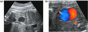
Purpose: Fetal intra-abdominal umbilical vein varix (FIUVV) can cause thrombosis, fetal growth restriction (FGR), and intrauterine fetal death (IUFD). However, its management and evaluation to avoid fetal risks have not been elucidated. The aim of this study was to develop a novel method to evaluate fetal risks, including FGR and fetal dysfunction via frequent ultrasound examinations.
Methods: A 28-year-old pregnant woman was diagnosed with FIUVV via ultrasound at 26 weeks of gestation and admitted to our hospital. Ultrasound examinations were performed two to three times weekly to evaluate size and shape of the FIUVV and umbilical vein blood flow at the inflow and outflow sites of the FIUVV.
Results: The outflow site of the FIUVV was constricted and collapsed, and the blood flow velocity at the inflow site of the FIUVV was decreased. At 32 weeks of gestation, spontaneous echo contrast (SEC), which indicates increased echogenicity, appeared. At 35 weeks of gestation, the patient noticed decreased fetal movement, and CTG showed non-reassuring fetal status. SEC in the FIUVV was remarkable. Fetal movement could not be confirmed at ultrasound. Cesarean section was performed and a 1,854-g healthy infant was delivered with an umbilical cord arterial pH of 7.266.
Conclusion: The echographic changes, such as decreased umbilical vein blood flow and SEC, in FIUVV observed in this case could indicate thrombus formation, which can lead to fetal dysfunction. Frequent ultrasound examinations can help determine the timing of delivery and improve the neonatal prognosis.
期刊介绍:
The Journal of Medical Ultrasonics is the official journal of the Japan Society of Ultrasonics in Medicine. The main purpose of the journal is to provide forum for the publication of papers documenting recent advances and new developments in the entire field of ultrasound in medicine and biology, encompassing both the medical and the engineering aspects of the science.The journal welcomes original articles, review articles, images, and letters to the editor.The journal also provides state-of-the-art information such as announcements from the boards and the committees of the society.


 分享
分享
 求助内容:
求助内容: 应助结果提醒方式:
 扫码关注我们
