{"title":"动脉自旋标记磁共振成像和神经认知及其他精神障碍的灌注模式:系统综述。","authors":"Rita Ferreira, António J Bastos-Leite","doi":"10.1007/s00234-024-03323-0","DOIUrl":null,"url":null,"abstract":"<p><p>We reviewed 33 original research studies assessing brain perfusion, using consensus guidelines from a \"white paper\" issued by the International Society for Magnetic Resonance in Medicine Perfusion Study Group and the European Cooperation in Science and Technology Action BM1103 (\"Arterial Spin Labelling Initiative in Dementia\"; https://www.cost.eu/actions/BM1103/ ). The studies were published between 2011 and 2023 and included participants with subjective cognitive decline plus; neurocognitive disorders, including mild cognitive impairment (MCI), Alzheimer's disease (AD), frontotemporal lobar degeneration (FTLD), dementia with Lewy bodies (DLB) and vascular cognitive impairment (VCI); as well as schizophrenia spectrum disorders, bipolar and major depressive disorders, autism spectrum disorder, attention-deficit/hyperactivity disorder, panic disorder and alcohol use disorder. Hypoperfusion associated with cognitive impairment was the major finding across the spectrum of cognitive decline. Regional hyperperfusion also was reported in MCI, AD, frontotemporal dementia phenocopy syndrome and VCI. Hypoperfused structures found to aid in diagnosing AD included the precunei and adjacent posterior cingulate cortices. Hypoperfused structures found to better diagnose patients with FTLD were the anterior cingulate cortices and frontal regions. Hypoperfusion in patients with DLB was found to relatively spare the temporal lobes, even after correction for partial volume effects. Hyperperfusion in the temporal cortices and hypoperfusion in the prefrontal and anterior cingulate cortices were found in patients with schizophrenia, most of whom were on medication and at the chronic stage of illness. Infratentorial structures were found to be abnormally perfused in patients with bipolar or major depressive disorders. Brain perfusion abnormalities were helpful in diagnosing most neurocognitive disorders. Abnormalities reported in VCI and the remaining mental disorders were heterogeneous and not generalisable.</p>","PeriodicalId":19422,"journal":{"name":"Neuroradiology","volume":" ","pages":"1065-1081"},"PeriodicalIF":2.6000,"publicationDate":"2024-07-01","publicationTypes":"Journal Article","fieldsOfStudy":null,"isOpenAccess":false,"openAccessPdf":"https://www.ncbi.nlm.nih.gov/pmc/articles/PMC11150205/pdf/","citationCount":"0","resultStr":"{\"title\":\"Arterial spin labelling magnetic resonance imaging and perfusion patterns in neurocognitive and other mental disorders: a systematic review.\",\"authors\":\"Rita Ferreira, António J Bastos-Leite\",\"doi\":\"10.1007/s00234-024-03323-0\",\"DOIUrl\":null,\"url\":null,\"abstract\":\"<p><p>We reviewed 33 original research studies assessing brain perfusion, using consensus guidelines from a \\\"white paper\\\" issued by the International Society for Magnetic Resonance in Medicine Perfusion Study Group and the European Cooperation in Science and Technology Action BM1103 (\\\"Arterial Spin Labelling Initiative in Dementia\\\"; https://www.cost.eu/actions/BM1103/ ). The studies were published between 2011 and 2023 and included participants with subjective cognitive decline plus; neurocognitive disorders, including mild cognitive impairment (MCI), Alzheimer's disease (AD), frontotemporal lobar degeneration (FTLD), dementia with Lewy bodies (DLB) and vascular cognitive impairment (VCI); as well as schizophrenia spectrum disorders, bipolar and major depressive disorders, autism spectrum disorder, attention-deficit/hyperactivity disorder, panic disorder and alcohol use disorder. Hypoperfusion associated with cognitive impairment was the major finding across the spectrum of cognitive decline. Regional hyperperfusion also was reported in MCI, AD, frontotemporal dementia phenocopy syndrome and VCI. Hypoperfused structures found to aid in diagnosing AD included the precunei and adjacent posterior cingulate cortices. Hypoperfused structures found to better diagnose patients with FTLD were the anterior cingulate cortices and frontal regions. Hypoperfusion in patients with DLB was found to relatively spare the temporal lobes, even after correction for partial volume effects. Hyperperfusion in the temporal cortices and hypoperfusion in the prefrontal and anterior cingulate cortices were found in patients with schizophrenia, most of whom were on medication and at the chronic stage of illness. Infratentorial structures were found to be abnormally perfused in patients with bipolar or major depressive disorders. Brain perfusion abnormalities were helpful in diagnosing most neurocognitive disorders. Abnormalities reported in VCI and the remaining mental disorders were heterogeneous and not generalisable.</p>\",\"PeriodicalId\":19422,\"journal\":{\"name\":\"Neuroradiology\",\"volume\":\" \",\"pages\":\"1065-1081\"},\"PeriodicalIF\":2.6000,\"publicationDate\":\"2024-07-01\",\"publicationTypes\":\"Journal Article\",\"fieldsOfStudy\":null,\"isOpenAccess\":false,\"openAccessPdf\":\"https://www.ncbi.nlm.nih.gov/pmc/articles/PMC11150205/pdf/\",\"citationCount\":\"0\",\"resultStr\":null,\"platform\":\"Semanticscholar\",\"paperid\":null,\"PeriodicalName\":\"Neuroradiology\",\"FirstCategoryId\":\"3\",\"ListUrlMain\":\"https://doi.org/10.1007/s00234-024-03323-0\",\"RegionNum\":3,\"RegionCategory\":\"医学\",\"ArticlePicture\":[],\"TitleCN\":null,\"AbstractTextCN\":null,\"PMCID\":null,\"EPubDate\":\"2024/3/27 0:00:00\",\"PubModel\":\"Epub\",\"JCR\":\"Q2\",\"JCRName\":\"CLINICAL NEUROLOGY\",\"Score\":null,\"Total\":0}","platform":"Semanticscholar","paperid":null,"PeriodicalName":"Neuroradiology","FirstCategoryId":"3","ListUrlMain":"https://doi.org/10.1007/s00234-024-03323-0","RegionNum":3,"RegionCategory":"医学","ArticlePicture":[],"TitleCN":null,"AbstractTextCN":null,"PMCID":null,"EPubDate":"2024/3/27 0:00:00","PubModel":"Epub","JCR":"Q2","JCRName":"CLINICAL NEUROLOGY","Score":null,"Total":0}
Arterial spin labelling magnetic resonance imaging and perfusion patterns in neurocognitive and other mental disorders: a systematic review.
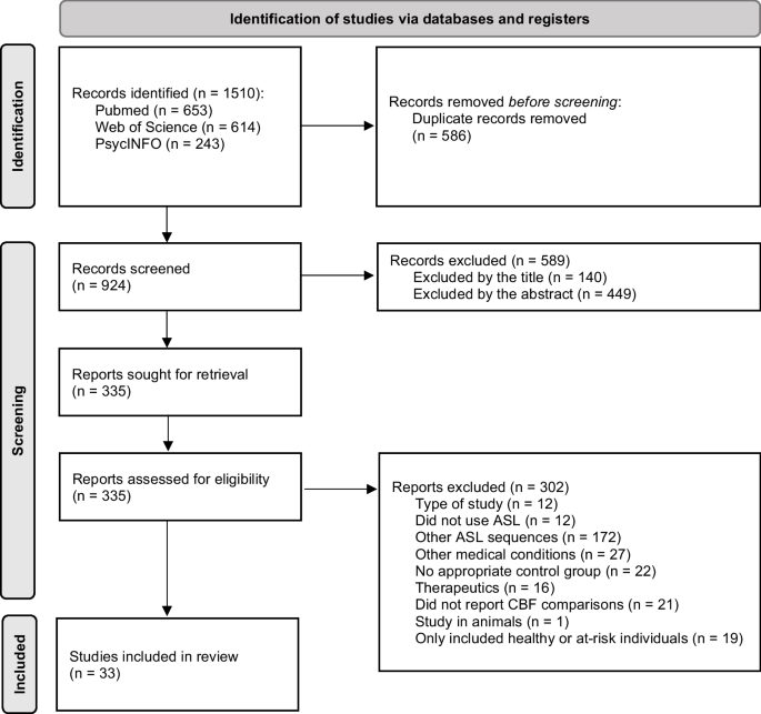
We reviewed 33 original research studies assessing brain perfusion, using consensus guidelines from a "white paper" issued by the International Society for Magnetic Resonance in Medicine Perfusion Study Group and the European Cooperation in Science and Technology Action BM1103 ("Arterial Spin Labelling Initiative in Dementia"; https://www.cost.eu/actions/BM1103/ ). The studies were published between 2011 and 2023 and included participants with subjective cognitive decline plus; neurocognitive disorders, including mild cognitive impairment (MCI), Alzheimer's disease (AD), frontotemporal lobar degeneration (FTLD), dementia with Lewy bodies (DLB) and vascular cognitive impairment (VCI); as well as schizophrenia spectrum disorders, bipolar and major depressive disorders, autism spectrum disorder, attention-deficit/hyperactivity disorder, panic disorder and alcohol use disorder. Hypoperfusion associated with cognitive impairment was the major finding across the spectrum of cognitive decline. Regional hyperperfusion also was reported in MCI, AD, frontotemporal dementia phenocopy syndrome and VCI. Hypoperfused structures found to aid in diagnosing AD included the precunei and adjacent posterior cingulate cortices. Hypoperfused structures found to better diagnose patients with FTLD were the anterior cingulate cortices and frontal regions. Hypoperfusion in patients with DLB was found to relatively spare the temporal lobes, even after correction for partial volume effects. Hyperperfusion in the temporal cortices and hypoperfusion in the prefrontal and anterior cingulate cortices were found in patients with schizophrenia, most of whom were on medication and at the chronic stage of illness. Infratentorial structures were found to be abnormally perfused in patients with bipolar or major depressive disorders. Brain perfusion abnormalities were helpful in diagnosing most neurocognitive disorders. Abnormalities reported in VCI and the remaining mental disorders were heterogeneous and not generalisable.
期刊介绍:
Neuroradiology aims to provide state-of-the-art medical and scientific information in the fields of Neuroradiology, Neurosciences, Neurology, Psychiatry, Neurosurgery, and related medical specialities. Neuroradiology as the official Journal of the European Society of Neuroradiology receives submissions from all parts of the world and publishes peer-reviewed original research, comprehensive reviews, educational papers, opinion papers, and short reports on exceptional clinical observations and new technical developments in the field of Neuroimaging and Neurointervention. The journal has subsections for Diagnostic and Interventional Neuroradiology, Advanced Neuroimaging, Paediatric Neuroradiology, Head-Neck-ENT Radiology, Spine Neuroradiology, and for submissions from Japan. Neuroradiology aims to provide new knowledge about and insights into the function and pathology of the human nervous system that may help to better diagnose and treat nervous system diseases. Neuroradiology is a member of the Committee on Publication Ethics (COPE) and follows the COPE core practices. Neuroradiology prefers articles that are free of bias, self-critical regarding limitations, transparent and clear in describing study participants, methods, and statistics, and short in presenting results. Before peer-review all submissions are automatically checked by iThenticate to assess for potential overlap in prior publication.


 分享
分享
 求助内容:
求助内容: 应助结果提醒方式:
 扫码关注我们
