Yaping Ju, Miriam Velazquez, Andy Sherrod, Tiannan Wang
{"title":"通过 EBUS-FNA 诊断 NUT 癌的诊断意义和细胞学特征,病例报告和文献综述。","authors":"Yaping Ju, Miriam Velazquez, Andy Sherrod, Tiannan Wang","doi":"10.1111/cyt.13379","DOIUrl":null,"url":null,"abstract":"<p>Cytomorphological features of NUT carcinoma include sheets or discrete nests of primitive, monotonous, round to oval shaped tumour cells with high N/C ratio and brisk mitotic figures. Abrupt squamous differentiation might be a diagnostic hint. More than 50% positivity of NUT immunohistochemistry staining is diagnostic.\n <figure>\n <div><picture>\n <source></source></picture><p></p>\n </div>\n </figure></p><p>NUT carcinoma represents a poorly differentiated malignancy by extremely aggressive clinical course and poor prognosis. It frequently manifests in midline organs, notably in the mediastinum and lung. The rising preferences for utilizing the EBUS-FNA procedure in diagnosing thoracic and lung lesions stems from its high diagnostic yield. Hence, recognizing the cytomorphological features of NUT carcinoma is crucial for timely treatment and improved patient survival.</p>","PeriodicalId":55187,"journal":{"name":"Cytopathology","volume":"35 4","pages":"497-502"},"PeriodicalIF":1.1000,"publicationDate":"2024-03-29","publicationTypes":"Journal Article","fieldsOfStudy":null,"isOpenAccess":false,"openAccessPdf":"https://onlinelibrary.wiley.com/doi/epdf/10.1111/cyt.13379","citationCount":"0","resultStr":"{\"title\":\"Diagnostic significance and cytological features of NUT carcinoma by EBUS-FNA, a case report and literature review\",\"authors\":\"Yaping Ju, Miriam Velazquez, Andy Sherrod, Tiannan Wang\",\"doi\":\"10.1111/cyt.13379\",\"DOIUrl\":null,\"url\":null,\"abstract\":\"<p>Cytomorphological features of NUT carcinoma include sheets or discrete nests of primitive, monotonous, round to oval shaped tumour cells with high N/C ratio and brisk mitotic figures. Abrupt squamous differentiation might be a diagnostic hint. More than 50% positivity of NUT immunohistochemistry staining is diagnostic.\\n <figure>\\n <div><picture>\\n <source></source></picture><p></p>\\n </div>\\n </figure></p><p>NUT carcinoma represents a poorly differentiated malignancy by extremely aggressive clinical course and poor prognosis. It frequently manifests in midline organs, notably in the mediastinum and lung. The rising preferences for utilizing the EBUS-FNA procedure in diagnosing thoracic and lung lesions stems from its high diagnostic yield. Hence, recognizing the cytomorphological features of NUT carcinoma is crucial for timely treatment and improved patient survival.</p>\",\"PeriodicalId\":55187,\"journal\":{\"name\":\"Cytopathology\",\"volume\":\"35 4\",\"pages\":\"497-502\"},\"PeriodicalIF\":1.1000,\"publicationDate\":\"2024-03-29\",\"publicationTypes\":\"Journal Article\",\"fieldsOfStudy\":null,\"isOpenAccess\":false,\"openAccessPdf\":\"https://onlinelibrary.wiley.com/doi/epdf/10.1111/cyt.13379\",\"citationCount\":\"0\",\"resultStr\":null,\"platform\":\"Semanticscholar\",\"paperid\":null,\"PeriodicalName\":\"Cytopathology\",\"FirstCategoryId\":\"3\",\"ListUrlMain\":\"https://onlinelibrary.wiley.com/doi/10.1111/cyt.13379\",\"RegionNum\":4,\"RegionCategory\":\"医学\",\"ArticlePicture\":[],\"TitleCN\":null,\"AbstractTextCN\":null,\"PMCID\":null,\"EPubDate\":\"\",\"PubModel\":\"\",\"JCR\":\"Q4\",\"JCRName\":\"CELL BIOLOGY\",\"Score\":null,\"Total\":0}","platform":"Semanticscholar","paperid":null,"PeriodicalName":"Cytopathology","FirstCategoryId":"3","ListUrlMain":"https://onlinelibrary.wiley.com/doi/10.1111/cyt.13379","RegionNum":4,"RegionCategory":"医学","ArticlePicture":[],"TitleCN":null,"AbstractTextCN":null,"PMCID":null,"EPubDate":"","PubModel":"","JCR":"Q4","JCRName":"CELL BIOLOGY","Score":null,"Total":0}
Diagnostic significance and cytological features of NUT carcinoma by EBUS-FNA, a case report and literature review
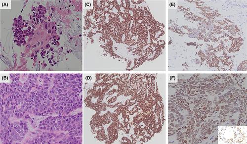
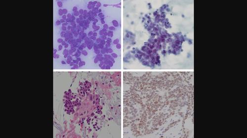
Cytomorphological features of NUT carcinoma include sheets or discrete nests of primitive, monotonous, round to oval shaped tumour cells with high N/C ratio and brisk mitotic figures. Abrupt squamous differentiation might be a diagnostic hint. More than 50% positivity of NUT immunohistochemistry staining is diagnostic.
NUT carcinoma represents a poorly differentiated malignancy by extremely aggressive clinical course and poor prognosis. It frequently manifests in midline organs, notably in the mediastinum and lung. The rising preferences for utilizing the EBUS-FNA procedure in diagnosing thoracic and lung lesions stems from its high diagnostic yield. Hence, recognizing the cytomorphological features of NUT carcinoma is crucial for timely treatment and improved patient survival.
期刊介绍:
The aim of Cytopathology is to publish articles relating to those aspects of cytology which will increase our knowledge and understanding of the aetiology, diagnosis and management of human disease. It contains original articles and critical reviews on all aspects of clinical cytology in its broadest sense, including: gynaecological and non-gynaecological cytology; fine needle aspiration and screening strategy.
Cytopathology welcomes papers and articles on: ultrastructural, histochemical and immunocytochemical studies of the cell; quantitative cytology and DNA hybridization as applied to cytological material.


 分享
分享
 求助内容:
求助内容: 应助结果提醒方式:
 扫码关注我们
