Dylan Haynes MD, MCR, Eckart Haneke MD, PhD, Adam I. Rubin MD
{"title":"恶性绒毛膜瘤的临床、虹膜镜检查、指甲剪切和组织病理学发现。","authors":"Dylan Haynes MD, MCR, Eckart Haneke MD, PhD, Adam I. Rubin MD","doi":"10.1111/cup.14620","DOIUrl":null,"url":null,"abstract":"<p>This report describes the clinical, onychoscopic, nail clipping, and histopathologic features of a malignant onychopapilloma. A 71-year-old male presented to our outpatient clinic for a stable, asymptomatic lesion on his left middle finger that had been present for 2 years. Prior nail clipping histopathology showed nail plate thinning with subungual abnormal onychocytes. Clinical examination revealed a 2-mm-wide streak of longitudinal xanthonychia extending to the proximal nail fold, with distal hyperkeratosis and onycholysis. Onychoscopy showed irregular longitudinal nail plate ridging with scattered punctate hemorrhagic foci. An excisional nail unit biopsy demonstrated cellular atypia of the nail bed epithelium, matrix metaplasia, longitudinal abnormal onychocytes, increased Ki-67 staining, and negative HPV immunoperoxidase staining, confirming the diagnosis of malignant onychopapilloma.</p>","PeriodicalId":15407,"journal":{"name":"Journal of Cutaneous Pathology","volume":null,"pages":null},"PeriodicalIF":1.6000,"publicationDate":"2024-04-02","publicationTypes":"Journal Article","fieldsOfStudy":null,"isOpenAccess":false,"openAccessPdf":"https://onlinelibrary.wiley.com/doi/epdf/10.1111/cup.14620","citationCount":"0","resultStr":"{\"title\":\"Clinical, onychoscopic, nail clipping, and histopathological findings of malignant onychopapilloma\",\"authors\":\"Dylan Haynes MD, MCR, Eckart Haneke MD, PhD, Adam I. Rubin MD\",\"doi\":\"10.1111/cup.14620\",\"DOIUrl\":null,\"url\":null,\"abstract\":\"<p>This report describes the clinical, onychoscopic, nail clipping, and histopathologic features of a malignant onychopapilloma. A 71-year-old male presented to our outpatient clinic for a stable, asymptomatic lesion on his left middle finger that had been present for 2 years. Prior nail clipping histopathology showed nail plate thinning with subungual abnormal onychocytes. Clinical examination revealed a 2-mm-wide streak of longitudinal xanthonychia extending to the proximal nail fold, with distal hyperkeratosis and onycholysis. Onychoscopy showed irregular longitudinal nail plate ridging with scattered punctate hemorrhagic foci. An excisional nail unit biopsy demonstrated cellular atypia of the nail bed epithelium, matrix metaplasia, longitudinal abnormal onychocytes, increased Ki-67 staining, and negative HPV immunoperoxidase staining, confirming the diagnosis of malignant onychopapilloma.</p>\",\"PeriodicalId\":15407,\"journal\":{\"name\":\"Journal of Cutaneous Pathology\",\"volume\":null,\"pages\":null},\"PeriodicalIF\":1.6000,\"publicationDate\":\"2024-04-02\",\"publicationTypes\":\"Journal Article\",\"fieldsOfStudy\":null,\"isOpenAccess\":false,\"openAccessPdf\":\"https://onlinelibrary.wiley.com/doi/epdf/10.1111/cup.14620\",\"citationCount\":\"0\",\"resultStr\":null,\"platform\":\"Semanticscholar\",\"paperid\":null,\"PeriodicalName\":\"Journal of Cutaneous Pathology\",\"FirstCategoryId\":\"3\",\"ListUrlMain\":\"https://onlinelibrary.wiley.com/doi/10.1111/cup.14620\",\"RegionNum\":4,\"RegionCategory\":\"医学\",\"ArticlePicture\":[],\"TitleCN\":null,\"AbstractTextCN\":null,\"PMCID\":null,\"EPubDate\":\"\",\"PubModel\":\"\",\"JCR\":\"Q3\",\"JCRName\":\"DERMATOLOGY\",\"Score\":null,\"Total\":0}","platform":"Semanticscholar","paperid":null,"PeriodicalName":"Journal of Cutaneous Pathology","FirstCategoryId":"3","ListUrlMain":"https://onlinelibrary.wiley.com/doi/10.1111/cup.14620","RegionNum":4,"RegionCategory":"医学","ArticlePicture":[],"TitleCN":null,"AbstractTextCN":null,"PMCID":null,"EPubDate":"","PubModel":"","JCR":"Q3","JCRName":"DERMATOLOGY","Score":null,"Total":0}
Clinical, onychoscopic, nail clipping, and histopathological findings of malignant onychopapilloma
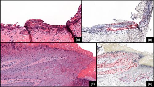
This report describes the clinical, onychoscopic, nail clipping, and histopathologic features of a malignant onychopapilloma. A 71-year-old male presented to our outpatient clinic for a stable, asymptomatic lesion on his left middle finger that had been present for 2 years. Prior nail clipping histopathology showed nail plate thinning with subungual abnormal onychocytes. Clinical examination revealed a 2-mm-wide streak of longitudinal xanthonychia extending to the proximal nail fold, with distal hyperkeratosis and onycholysis. Onychoscopy showed irregular longitudinal nail plate ridging with scattered punctate hemorrhagic foci. An excisional nail unit biopsy demonstrated cellular atypia of the nail bed epithelium, matrix metaplasia, longitudinal abnormal onychocytes, increased Ki-67 staining, and negative HPV immunoperoxidase staining, confirming the diagnosis of malignant onychopapilloma.
期刊介绍:
Journal of Cutaneous Pathology publishes manuscripts broadly relevant to diseases of the skin and mucosae, with the aims of advancing scientific knowledge regarding dermatopathology and enhancing the communication between clinical practitioners and research scientists. Original scientific manuscripts on diagnostic and experimental cutaneous pathology are especially desirable. Timely, pertinent review articles also will be given high priority. Manuscripts based on light, fluorescence, and electron microscopy, histochemistry, immunology, molecular biology, and genetics, as well as allied sciences, are all welcome, provided their principal focus is on cutaneous pathology. Publication time will be kept as short as possible, ensuring that articles will be quickly available to all interested in this speciality.


 分享
分享
 求助内容:
求助内容: 应助结果提醒方式:
 扫码关注我们
