{"title":"利用肺泡模型研究呼吸道致敏物质的转录组特征","authors":"Matthew Gibb, James Y. Liu, Christie M. Sayes","doi":"10.1007/s10565-024-09860-x","DOIUrl":null,"url":null,"abstract":"<p>Environmental contaminants are ubiquitous in the air we breathe and can potentially cause adverse immunological outcomes such as respiratory sensitization, a type of immune-driven allergic response in the lungs. Wood dust, latex, pet dander, oils, fragrances, paints, and glues have all been implicated as possible respiratory sensitizers. With the increased incidence of exposure to chemical mixtures and the rapid production of novel materials, it is paramount that testing regimes accounting for sensitization are incorporated into development cycles. However, no validated assay exists that is universally accepted to measure a substance’s respiratory sensitizing potential. The lungs comprise various cell types and regions where sensitization can occur, with the gas-exchange interface being especially important due to implications for overall lung function. As such, an assay that can mimic the alveolar compartment and assess sensitization would be an important advance for inhalation toxicology. Some such models are under development, but in-depth transcriptomic analyses have yet to be reported. Understanding the transcriptome after sensitizer exposure would greatly advance hazard assessment and sustainability. We tested two known sensitizers (<i>i.e.,</i> isophorone diisocyanate and ethylenediamine) and two known non-sensitizers (<i>i.e.,</i> chlorobenzene and dimethylformamide). RNA sequencing was performed in our in vitro alveolar model, consisting of a 3D co-culture of epithelial, macrophage, and dendritic cells. Sensitizers were readily distinguishable from non-sensitizers by principal component analysis. However, few differentially regulated genes were common across all pair-wise comparisons (<i>i.e.,</i> upregulation of genes <i>SOX9</i>, <i>UACA</i>, <i>CCDC88A</i>, <i>FOSL1</i>, <i>KIF20B</i>). While the model utilized in this study can differentiate the sensitizers from the non-sensitizers tested, further studies will be required to robustly identify critical pathways inducing respiratory sensitization.</p><h3 data-test=\"abstract-sub-heading\">Graphical Abstract</h3><p>Graphical headlines/headlights</p><ul>\n<li>\n<p>Pollutants may trigger lung allergies, but no universal method measures respiratory sensitization potential.</p>\n</li>\n<li>\n<p>In vitro systems can detect respiratory sensitizers, aiding in anticipating and reducing the risks of new materials.</p>\n</li>\n<li>\n<p>Sensitizers and non-sensitizers can be distinguished through transcriptome investigation.</p>\n</li>\n<li>\n<p>The sensitizers tested induced cell differentiation and proliferation pathways while inhibiting immune defense and functionality.</p>\n</li>\n</ul>\n","PeriodicalId":9672,"journal":{"name":"Cell Biology and Toxicology","volume":"41 1","pages":""},"PeriodicalIF":5.9000,"publicationDate":"2024-04-08","publicationTypes":"Journal Article","fieldsOfStudy":null,"isOpenAccess":false,"openAccessPdf":"","citationCount":"0","resultStr":"{\"title\":\"The transcriptomic signature of respiratory sensitizers using an alveolar model\",\"authors\":\"Matthew Gibb, James Y. Liu, Christie M. Sayes\",\"doi\":\"10.1007/s10565-024-09860-x\",\"DOIUrl\":null,\"url\":null,\"abstract\":\"<p>Environmental contaminants are ubiquitous in the air we breathe and can potentially cause adverse immunological outcomes such as respiratory sensitization, a type of immune-driven allergic response in the lungs. Wood dust, latex, pet dander, oils, fragrances, paints, and glues have all been implicated as possible respiratory sensitizers. With the increased incidence of exposure to chemical mixtures and the rapid production of novel materials, it is paramount that testing regimes accounting for sensitization are incorporated into development cycles. However, no validated assay exists that is universally accepted to measure a substance’s respiratory sensitizing potential. The lungs comprise various cell types and regions where sensitization can occur, with the gas-exchange interface being especially important due to implications for overall lung function. As such, an assay that can mimic the alveolar compartment and assess sensitization would be an important advance for inhalation toxicology. Some such models are under development, but in-depth transcriptomic analyses have yet to be reported. Understanding the transcriptome after sensitizer exposure would greatly advance hazard assessment and sustainability. We tested two known sensitizers (<i>i.e.,</i> isophorone diisocyanate and ethylenediamine) and two known non-sensitizers (<i>i.e.,</i> chlorobenzene and dimethylformamide). RNA sequencing was performed in our in vitro alveolar model, consisting of a 3D co-culture of epithelial, macrophage, and dendritic cells. Sensitizers were readily distinguishable from non-sensitizers by principal component analysis. However, few differentially regulated genes were common across all pair-wise comparisons (<i>i.e.,</i> upregulation of genes <i>SOX9</i>, <i>UACA</i>, <i>CCDC88A</i>, <i>FOSL1</i>, <i>KIF20B</i>). While the model utilized in this study can differentiate the sensitizers from the non-sensitizers tested, further studies will be required to robustly identify critical pathways inducing respiratory sensitization.</p><h3 data-test=\\\"abstract-sub-heading\\\">Graphical Abstract</h3><p>Graphical headlines/headlights</p><ul>\\n<li>\\n<p>Pollutants may trigger lung allergies, but no universal method measures respiratory sensitization potential.</p>\\n</li>\\n<li>\\n<p>In vitro systems can detect respiratory sensitizers, aiding in anticipating and reducing the risks of new materials.</p>\\n</li>\\n<li>\\n<p>Sensitizers and non-sensitizers can be distinguished through transcriptome investigation.</p>\\n</li>\\n<li>\\n<p>The sensitizers tested induced cell differentiation and proliferation pathways while inhibiting immune defense and functionality.</p>\\n</li>\\n</ul>\\n\",\"PeriodicalId\":9672,\"journal\":{\"name\":\"Cell Biology and Toxicology\",\"volume\":\"41 1\",\"pages\":\"\"},\"PeriodicalIF\":5.9000,\"publicationDate\":\"2024-04-08\",\"publicationTypes\":\"Journal Article\",\"fieldsOfStudy\":null,\"isOpenAccess\":false,\"openAccessPdf\":\"\",\"citationCount\":\"0\",\"resultStr\":null,\"platform\":\"Semanticscholar\",\"paperid\":null,\"PeriodicalName\":\"Cell Biology and Toxicology\",\"FirstCategoryId\":\"3\",\"ListUrlMain\":\"https://doi.org/10.1007/s10565-024-09860-x\",\"RegionNum\":2,\"RegionCategory\":\"医学\",\"ArticlePicture\":[],\"TitleCN\":null,\"AbstractTextCN\":null,\"PMCID\":null,\"EPubDate\":\"\",\"PubModel\":\"\",\"JCR\":\"Q2\",\"JCRName\":\"CELL BIOLOGY\",\"Score\":null,\"Total\":0}","platform":"Semanticscholar","paperid":null,"PeriodicalName":"Cell Biology and Toxicology","FirstCategoryId":"3","ListUrlMain":"https://doi.org/10.1007/s10565-024-09860-x","RegionNum":2,"RegionCategory":"医学","ArticlePicture":[],"TitleCN":null,"AbstractTextCN":null,"PMCID":null,"EPubDate":"","PubModel":"","JCR":"Q2","JCRName":"CELL BIOLOGY","Score":null,"Total":0}
The transcriptomic signature of respiratory sensitizers using an alveolar model
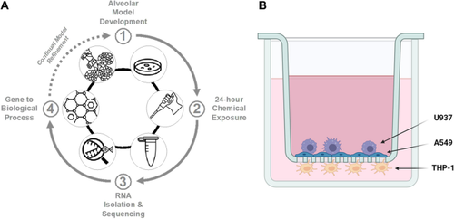
Environmental contaminants are ubiquitous in the air we breathe and can potentially cause adverse immunological outcomes such as respiratory sensitization, a type of immune-driven allergic response in the lungs. Wood dust, latex, pet dander, oils, fragrances, paints, and glues have all been implicated as possible respiratory sensitizers. With the increased incidence of exposure to chemical mixtures and the rapid production of novel materials, it is paramount that testing regimes accounting for sensitization are incorporated into development cycles. However, no validated assay exists that is universally accepted to measure a substance’s respiratory sensitizing potential. The lungs comprise various cell types and regions where sensitization can occur, with the gas-exchange interface being especially important due to implications for overall lung function. As such, an assay that can mimic the alveolar compartment and assess sensitization would be an important advance for inhalation toxicology. Some such models are under development, but in-depth transcriptomic analyses have yet to be reported. Understanding the transcriptome after sensitizer exposure would greatly advance hazard assessment and sustainability. We tested two known sensitizers (i.e., isophorone diisocyanate and ethylenediamine) and two known non-sensitizers (i.e., chlorobenzene and dimethylformamide). RNA sequencing was performed in our in vitro alveolar model, consisting of a 3D co-culture of epithelial, macrophage, and dendritic cells. Sensitizers were readily distinguishable from non-sensitizers by principal component analysis. However, few differentially regulated genes were common across all pair-wise comparisons (i.e., upregulation of genes SOX9, UACA, CCDC88A, FOSL1, KIF20B). While the model utilized in this study can differentiate the sensitizers from the non-sensitizers tested, further studies will be required to robustly identify critical pathways inducing respiratory sensitization.
Graphical Abstract
Graphical headlines/headlights
Pollutants may trigger lung allergies, but no universal method measures respiratory sensitization potential.
In vitro systems can detect respiratory sensitizers, aiding in anticipating and reducing the risks of new materials.
Sensitizers and non-sensitizers can be distinguished through transcriptome investigation.
The sensitizers tested induced cell differentiation and proliferation pathways while inhibiting immune defense and functionality.
期刊介绍:
Cell Biology and Toxicology (CBT) is an international journal focused on clinical and translational research with an emphasis on molecular and cell biology, genetic and epigenetic heterogeneity, drug discovery and development, and molecular pharmacology and toxicology. CBT has a disease-specific scope prioritizing publications on gene and protein-based regulation, intracellular signaling pathway dysfunction, cell type-specific function, and systems in biomedicine in drug discovery and development. CBT publishes original articles with outstanding, innovative and significant findings, important reviews on recent research advances and issues of high current interest, opinion articles of leading edge science, and rapid communication or reports, on molecular mechanisms and therapies in diseases.


 分享
分享
 求助内容:
求助内容: 应助结果提醒方式:
 扫码关注我们
