Ana Beatriz Raposo Souza, Adriana Dibo Cruz, Marcelo Freitas Aguiar
{"title":"利用锥形束计算机断层扫描对牙齿进行容积分析以估算年龄","authors":"Ana Beatriz Raposo Souza, Adriana Dibo Cruz, Marcelo Freitas Aguiar","doi":"10.1007/s11282-024-00750-w","DOIUrl":null,"url":null,"abstract":"<h3 data-test=\"abstract-sub-heading\">Objectives</h3><p>To evaluate the feasibility of using the pulp volume (Pv) to total volume (Tv) ratio (Pv:Tv), obtained from cone beam computed tomography (CBCT) scans of single-rooted teeth, for age estimation in a Brazilian population sample.</p><h3 data-test=\"abstract-sub-heading\">Methods</h3><p>After obtaining approval from the ethics committee, the study commenced by applying inclusion criteria to screen CBCT scans, resulting in a probability-based sample of participants aged 18 years and older (ranging from 18 to 82 years, with a mean age of 46.44 years). A total of 517 single-rooted teeth, including maxillary central incisors (CI), mandibular canines (C), and mandibular first premolars (FP), were chosen based on excellent agreement values (> 0.9). Pv and Tv measurements were conducted using semi-automatic segmentation with ITK-SNAP 3.8 software. Statistical analysis was performed using Jamovi software, with a significance level set at 5% (<i>α</i> = 0.05).</p><h3 data-test=\"abstract-sub-heading\">Results</h3><p>A strong negative correlation (<i>r</i> > −0.7) was observed between chronological age and the Pv:Tv ratio across all examined teeth. However, when conducting regression analysis with Pv:Tv data and chronological age as the independent variable, only the mandibular FP teeth exhibited a normal distribution. The resulting linear model demonstrated moderate predictive value (approximately 64%) in explaining the variance in chronological age, but caution should be exercised when interpreting these findings.</p><h3 data-test=\"abstract-sub-heading\">Conclusions</h3><p>The method of measuring individual tooth volume using CBCT to estimate chronological age via Pv:Tv has been demonstrated as effective and reproducible within the Brazilian population sample.</p>","PeriodicalId":56103,"journal":{"name":"Oral Radiology","volume":"11 1","pages":""},"PeriodicalIF":1.7000,"publicationDate":"2024-04-08","publicationTypes":"Journal Article","fieldsOfStudy":null,"isOpenAccess":false,"openAccessPdf":"","citationCount":"0","resultStr":"{\"title\":\"Age estimation by volumetric analysis of teeth using cone beam computed tomography\",\"authors\":\"Ana Beatriz Raposo Souza, Adriana Dibo Cruz, Marcelo Freitas Aguiar\",\"doi\":\"10.1007/s11282-024-00750-w\",\"DOIUrl\":null,\"url\":null,\"abstract\":\"<h3 data-test=\\\"abstract-sub-heading\\\">Objectives</h3><p>To evaluate the feasibility of using the pulp volume (Pv) to total volume (Tv) ratio (Pv:Tv), obtained from cone beam computed tomography (CBCT) scans of single-rooted teeth, for age estimation in a Brazilian population sample.</p><h3 data-test=\\\"abstract-sub-heading\\\">Methods</h3><p>After obtaining approval from the ethics committee, the study commenced by applying inclusion criteria to screen CBCT scans, resulting in a probability-based sample of participants aged 18 years and older (ranging from 18 to 82 years, with a mean age of 46.44 years). A total of 517 single-rooted teeth, including maxillary central incisors (CI), mandibular canines (C), and mandibular first premolars (FP), were chosen based on excellent agreement values (> 0.9). Pv and Tv measurements were conducted using semi-automatic segmentation with ITK-SNAP 3.8 software. Statistical analysis was performed using Jamovi software, with a significance level set at 5% (<i>α</i> = 0.05).</p><h3 data-test=\\\"abstract-sub-heading\\\">Results</h3><p>A strong negative correlation (<i>r</i> > −0.7) was observed between chronological age and the Pv:Tv ratio across all examined teeth. However, when conducting regression analysis with Pv:Tv data and chronological age as the independent variable, only the mandibular FP teeth exhibited a normal distribution. The resulting linear model demonstrated moderate predictive value (approximately 64%) in explaining the variance in chronological age, but caution should be exercised when interpreting these findings.</p><h3 data-test=\\\"abstract-sub-heading\\\">Conclusions</h3><p>The method of measuring individual tooth volume using CBCT to estimate chronological age via Pv:Tv has been demonstrated as effective and reproducible within the Brazilian population sample.</p>\",\"PeriodicalId\":56103,\"journal\":{\"name\":\"Oral Radiology\",\"volume\":\"11 1\",\"pages\":\"\"},\"PeriodicalIF\":1.7000,\"publicationDate\":\"2024-04-08\",\"publicationTypes\":\"Journal Article\",\"fieldsOfStudy\":null,\"isOpenAccess\":false,\"openAccessPdf\":\"\",\"citationCount\":\"0\",\"resultStr\":null,\"platform\":\"Semanticscholar\",\"paperid\":null,\"PeriodicalName\":\"Oral Radiology\",\"FirstCategoryId\":\"3\",\"ListUrlMain\":\"https://doi.org/10.1007/s11282-024-00750-w\",\"RegionNum\":3,\"RegionCategory\":\"医学\",\"ArticlePicture\":[],\"TitleCN\":null,\"AbstractTextCN\":null,\"PMCID\":null,\"EPubDate\":\"\",\"PubModel\":\"\",\"JCR\":\"Q3\",\"JCRName\":\"DENTISTRY, ORAL SURGERY & MEDICINE\",\"Score\":null,\"Total\":0}","platform":"Semanticscholar","paperid":null,"PeriodicalName":"Oral Radiology","FirstCategoryId":"3","ListUrlMain":"https://doi.org/10.1007/s11282-024-00750-w","RegionNum":3,"RegionCategory":"医学","ArticlePicture":[],"TitleCN":null,"AbstractTextCN":null,"PMCID":null,"EPubDate":"","PubModel":"","JCR":"Q3","JCRName":"DENTISTRY, ORAL SURGERY & MEDICINE","Score":null,"Total":0}
Age estimation by volumetric analysis of teeth using cone beam computed tomography
Objectives
To evaluate the feasibility of using the pulp volume (Pv) to total volume (Tv) ratio (Pv:Tv), obtained from cone beam computed tomography (CBCT) scans of single-rooted teeth, for age estimation in a Brazilian population sample.
Methods
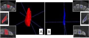
After obtaining approval from the ethics committee, the study commenced by applying inclusion criteria to screen CBCT scans, resulting in a probability-based sample of participants aged 18 years and older (ranging from 18 to 82 years, with a mean age of 46.44 years). A total of 517 single-rooted teeth, including maxillary central incisors (CI), mandibular canines (C), and mandibular first premolars (FP), were chosen based on excellent agreement values (> 0.9). Pv and Tv measurements were conducted using semi-automatic segmentation with ITK-SNAP 3.8 software. Statistical analysis was performed using Jamovi software, with a significance level set at 5% (α = 0.05).
Results
A strong negative correlation (r > −0.7) was observed between chronological age and the Pv:Tv ratio across all examined teeth. However, when conducting regression analysis with Pv:Tv data and chronological age as the independent variable, only the mandibular FP teeth exhibited a normal distribution. The resulting linear model demonstrated moderate predictive value (approximately 64%) in explaining the variance in chronological age, but caution should be exercised when interpreting these findings.
Conclusions
The method of measuring individual tooth volume using CBCT to estimate chronological age via Pv:Tv has been demonstrated as effective and reproducible within the Brazilian population sample.
期刊介绍:
As the official English-language journal of the Japanese Society for Oral and Maxillofacial Radiology and the Asian Academy of Oral and Maxillofacial Radiology, Oral Radiology is intended to be a forum for international collaboration in head and neck diagnostic imaging and all related fields. Oral Radiology features cutting-edge research papers, review articles, case reports, and technical notes from both the clinical and experimental fields. As membership in the Society is not a prerequisite, contributions are welcome from researchers and clinicians worldwide.


 分享
分享
 求助内容:
求助内容: 应助结果提醒方式:
 扫码关注我们
