Naoko Mori, Li Li, Masazumi Matsuda, Yu Mori, Shunji Mugikura
{"title":"灌注对比增强超声(CE-US)诊断乳腺癌腋窝淋巴结转移的前景:与淋巴CE-US的比较","authors":"Naoko Mori, Li Li, Masazumi Matsuda, Yu Mori, Shunji Mugikura","doi":"10.1007/s10396-024-01444-w","DOIUrl":null,"url":null,"abstract":"<p>Accurate diagnosis of lymph node (LN) metastasis is vital for prognosis and treatment in patients with breast cancer. Imaging 1modalities such as ultrasound (US), MRI, CT, and 18F-FDG PET/CT are used for preoperative assessment. While conventional US is commonly recommended due to its resolution and sensitivity, it has limitations such as operator subjectivity and difficulty detecting small metastases. This review shows the microanatomy of axillary LNs to enhance accurate diagnosis and the characteristics of contrast-enhanced US (CE-US), which utilizes intravascular microbubble contrast agents, making it ideal for vascular imaging. A significant focus of this review is on distinguishing between two types of CE-US techniques for axillary LN evaluation: perfusion CE-US and lymphatic CE-US. Perfusion CE-US is used to assess LN metastasis via transvenous contrast agent administration, while lymphatic CE-US is used to identify sentinel LNs and diagnose LN metastasis through percutaneous contrast agent administration. This review also highlights the need for future research to clarify the distinction between studies involving “apparently enlarged LNs” and “clinical node-negative” cases in perfusion CE-US research. Such research standardization is essential to ensure accurate diagnostic performance in various clinical studies. Future studies should aim to standardize CE-US methods for improved LN metastasis diagnosis, not only in breast cancer but also across various malignancies.</p>","PeriodicalId":50130,"journal":{"name":"Journal of Medical Ultrasonics","volume":"8 1","pages":""},"PeriodicalIF":2.1000,"publicationDate":"2024-04-20","publicationTypes":"Journal Article","fieldsOfStudy":null,"isOpenAccess":false,"openAccessPdf":"","citationCount":"0","resultStr":"{\"title\":\"Prospects of perfusion contrast-enhanced ultrasound (CE-US) in diagnosing axillary lymph node metastases in breast cancer: a comparison with lymphatic CE-US\",\"authors\":\"Naoko Mori, Li Li, Masazumi Matsuda, Yu Mori, Shunji Mugikura\",\"doi\":\"10.1007/s10396-024-01444-w\",\"DOIUrl\":null,\"url\":null,\"abstract\":\"<p>Accurate diagnosis of lymph node (LN) metastasis is vital for prognosis and treatment in patients with breast cancer. Imaging 1modalities such as ultrasound (US), MRI, CT, and 18F-FDG PET/CT are used for preoperative assessment. While conventional US is commonly recommended due to its resolution and sensitivity, it has limitations such as operator subjectivity and difficulty detecting small metastases. This review shows the microanatomy of axillary LNs to enhance accurate diagnosis and the characteristics of contrast-enhanced US (CE-US), which utilizes intravascular microbubble contrast agents, making it ideal for vascular imaging. A significant focus of this review is on distinguishing between two types of CE-US techniques for axillary LN evaluation: perfusion CE-US and lymphatic CE-US. Perfusion CE-US is used to assess LN metastasis via transvenous contrast agent administration, while lymphatic CE-US is used to identify sentinel LNs and diagnose LN metastasis through percutaneous contrast agent administration. This review also highlights the need for future research to clarify the distinction between studies involving “apparently enlarged LNs” and “clinical node-negative” cases in perfusion CE-US research. Such research standardization is essential to ensure accurate diagnostic performance in various clinical studies. Future studies should aim to standardize CE-US methods for improved LN metastasis diagnosis, not only in breast cancer but also across various malignancies.</p>\",\"PeriodicalId\":50130,\"journal\":{\"name\":\"Journal of Medical Ultrasonics\",\"volume\":\"8 1\",\"pages\":\"\"},\"PeriodicalIF\":2.1000,\"publicationDate\":\"2024-04-20\",\"publicationTypes\":\"Journal Article\",\"fieldsOfStudy\":null,\"isOpenAccess\":false,\"openAccessPdf\":\"\",\"citationCount\":\"0\",\"resultStr\":null,\"platform\":\"Semanticscholar\",\"paperid\":null,\"PeriodicalName\":\"Journal of Medical Ultrasonics\",\"FirstCategoryId\":\"3\",\"ListUrlMain\":\"https://doi.org/10.1007/s10396-024-01444-w\",\"RegionNum\":4,\"RegionCategory\":\"医学\",\"ArticlePicture\":[],\"TitleCN\":null,\"AbstractTextCN\":null,\"PMCID\":null,\"EPubDate\":\"\",\"PubModel\":\"\",\"JCR\":\"Q3\",\"JCRName\":\"RADIOLOGY, NUCLEAR MEDICINE & MEDICAL IMAGING\",\"Score\":null,\"Total\":0}","platform":"Semanticscholar","paperid":null,"PeriodicalName":"Journal of Medical Ultrasonics","FirstCategoryId":"3","ListUrlMain":"https://doi.org/10.1007/s10396-024-01444-w","RegionNum":4,"RegionCategory":"医学","ArticlePicture":[],"TitleCN":null,"AbstractTextCN":null,"PMCID":null,"EPubDate":"","PubModel":"","JCR":"Q3","JCRName":"RADIOLOGY, NUCLEAR MEDICINE & MEDICAL IMAGING","Score":null,"Total":0}
Prospects of perfusion contrast-enhanced ultrasound (CE-US) in diagnosing axillary lymph node metastases in breast cancer: a comparison with lymphatic CE-US
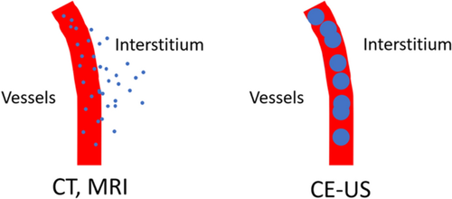
Accurate diagnosis of lymph node (LN) metastasis is vital for prognosis and treatment in patients with breast cancer. Imaging 1modalities such as ultrasound (US), MRI, CT, and 18F-FDG PET/CT are used for preoperative assessment. While conventional US is commonly recommended due to its resolution and sensitivity, it has limitations such as operator subjectivity and difficulty detecting small metastases. This review shows the microanatomy of axillary LNs to enhance accurate diagnosis and the characteristics of contrast-enhanced US (CE-US), which utilizes intravascular microbubble contrast agents, making it ideal for vascular imaging. A significant focus of this review is on distinguishing between two types of CE-US techniques for axillary LN evaluation: perfusion CE-US and lymphatic CE-US. Perfusion CE-US is used to assess LN metastasis via transvenous contrast agent administration, while lymphatic CE-US is used to identify sentinel LNs and diagnose LN metastasis through percutaneous contrast agent administration. This review also highlights the need for future research to clarify the distinction between studies involving “apparently enlarged LNs” and “clinical node-negative” cases in perfusion CE-US research. Such research standardization is essential to ensure accurate diagnostic performance in various clinical studies. Future studies should aim to standardize CE-US methods for improved LN metastasis diagnosis, not only in breast cancer but also across various malignancies.
期刊介绍:
The Journal of Medical Ultrasonics is the official journal of the Japan Society of Ultrasonics in Medicine. The main purpose of the journal is to provide forum for the publication of papers documenting recent advances and new developments in the entire field of ultrasound in medicine and biology, encompassing both the medical and the engineering aspects of the science.The journal welcomes original articles, review articles, images, and letters to the editor.The journal also provides state-of-the-art information such as announcements from the boards and the committees of the society.


 分享
分享
 求助内容:
求助内容: 应助结果提醒方式:
 扫码关注我们
