Soroush Irandoust , R. Christopher Whitton , Peter Muir , Corinne R. Henak
{"title":"纯血赛马第三掌骨髁旁沟槽中的软骨下骨疲劳损伤会提高特定部位的应变浓度","authors":"Soroush Irandoust , R. Christopher Whitton , Peter Muir , Corinne R. Henak","doi":"10.1016/j.jmbbm.2024.106561","DOIUrl":null,"url":null,"abstract":"<div><p>Condylar stress fracture of the distal end of the third metacarpal/metatarsal (MC3/MT3) bones is a major cause of Thoroughbred racehorse injury and euthanasia worldwide. Functional adaptation to exercise and fatigue damage lead to structural changes in the subchondral bone that include increased modeling (resulting in sclerotic bone tissue) and targeted remodeling repair (resulting in focal resorption spaces in the parasagittal groove). Whether these focal structural changes, as detectable by standing computed tomography (sCT), lead to elevated strain at the common site of condylar stress fracture has not been demonstrated. Therefore, the goal of the present study was to compare full-field three-dimensional (3D) strain on the distopalmar aspect of MC3 bone specimens with and without focal subchondral bone injury (SBI). Thirteen forelimb specimens were collected from racing Thoroughbreds for mechanical testing <em>ex vivo</em> and underwent sCT. Subsequently, full-field displacement and strain at the joint surface were determined using stereo digital image correlation. Strain concentration was observed in the parasagittal groove (PSG) of the loaded condyles, and those with SBI in the PSG showed higher strain rates in this region than control bones. PSG strain rate in condyles with PSG SBI was more sensitive to CT density distribution in comparison with condyles with no sCT-detectable injury. Findings from this study help to interpret structural changes in the subchondral bone due to fatigue damage and to assess risk of incipient stress fracture in a patient-specific manner.</p></div>","PeriodicalId":380,"journal":{"name":"Journal of the Mechanical Behavior of Biomedical Materials","volume":"155 ","pages":"Article 106561"},"PeriodicalIF":4.1000,"publicationDate":"2024-07-01","publicationTypes":"Journal Article","fieldsOfStudy":null,"isOpenAccess":false,"openAccessPdf":"","citationCount":"0","resultStr":"{\"title\":\"Subchondral bone fatigue injury in the parasagittal condylar grooves of the third metacarpal bone in thoroughbred racehorses elevates site-specific strain concentration\",\"authors\":\"Soroush Irandoust , R. Christopher Whitton , Peter Muir , Corinne R. Henak\",\"doi\":\"10.1016/j.jmbbm.2024.106561\",\"DOIUrl\":null,\"url\":null,\"abstract\":\"<div><p>Condylar stress fracture of the distal end of the third metacarpal/metatarsal (MC3/MT3) bones is a major cause of Thoroughbred racehorse injury and euthanasia worldwide. Functional adaptation to exercise and fatigue damage lead to structural changes in the subchondral bone that include increased modeling (resulting in sclerotic bone tissue) and targeted remodeling repair (resulting in focal resorption spaces in the parasagittal groove). Whether these focal structural changes, as detectable by standing computed tomography (sCT), lead to elevated strain at the common site of condylar stress fracture has not been demonstrated. Therefore, the goal of the present study was to compare full-field three-dimensional (3D) strain on the distopalmar aspect of MC3 bone specimens with and without focal subchondral bone injury (SBI). Thirteen forelimb specimens were collected from racing Thoroughbreds for mechanical testing <em>ex vivo</em> and underwent sCT. Subsequently, full-field displacement and strain at the joint surface were determined using stereo digital image correlation. Strain concentration was observed in the parasagittal groove (PSG) of the loaded condyles, and those with SBI in the PSG showed higher strain rates in this region than control bones. PSG strain rate in condyles with PSG SBI was more sensitive to CT density distribution in comparison with condyles with no sCT-detectable injury. Findings from this study help to interpret structural changes in the subchondral bone due to fatigue damage and to assess risk of incipient stress fracture in a patient-specific manner.</p></div>\",\"PeriodicalId\":380,\"journal\":{\"name\":\"Journal of the Mechanical Behavior of Biomedical Materials\",\"volume\":\"155 \",\"pages\":\"Article 106561\"},\"PeriodicalIF\":4.1000,\"publicationDate\":\"2024-07-01\",\"publicationTypes\":\"Journal Article\",\"fieldsOfStudy\":null,\"isOpenAccess\":false,\"openAccessPdf\":\"\",\"citationCount\":\"0\",\"resultStr\":null,\"platform\":\"Semanticscholar\",\"paperid\":null,\"PeriodicalName\":\"Journal of the Mechanical Behavior of Biomedical Materials\",\"FirstCategoryId\":\"5\",\"ListUrlMain\":\"https://www.sciencedirect.com/science/article/pii/S1751616124001930\",\"RegionNum\":2,\"RegionCategory\":\"医学\",\"ArticlePicture\":[],\"TitleCN\":null,\"AbstractTextCN\":null,\"PMCID\":null,\"EPubDate\":\"2024/4/24 0:00:00\",\"PubModel\":\"Epub\",\"JCR\":\"Q2\",\"JCRName\":\"ENGINEERING, BIOMEDICAL\",\"Score\":null,\"Total\":0}","platform":"Semanticscholar","paperid":null,"PeriodicalName":"Journal of the Mechanical Behavior of Biomedical Materials","FirstCategoryId":"5","ListUrlMain":"https://www.sciencedirect.com/science/article/pii/S1751616124001930","RegionNum":2,"RegionCategory":"医学","ArticlePicture":[],"TitleCN":null,"AbstractTextCN":null,"PMCID":null,"EPubDate":"2024/4/24 0:00:00","PubModel":"Epub","JCR":"Q2","JCRName":"ENGINEERING, BIOMEDICAL","Score":null,"Total":0}
Subchondral bone fatigue injury in the parasagittal condylar grooves of the third metacarpal bone in thoroughbred racehorses elevates site-specific strain concentration
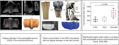
Condylar stress fracture of the distal end of the third metacarpal/metatarsal (MC3/MT3) bones is a major cause of Thoroughbred racehorse injury and euthanasia worldwide. Functional adaptation to exercise and fatigue damage lead to structural changes in the subchondral bone that include increased modeling (resulting in sclerotic bone tissue) and targeted remodeling repair (resulting in focal resorption spaces in the parasagittal groove). Whether these focal structural changes, as detectable by standing computed tomography (sCT), lead to elevated strain at the common site of condylar stress fracture has not been demonstrated. Therefore, the goal of the present study was to compare full-field three-dimensional (3D) strain on the distopalmar aspect of MC3 bone specimens with and without focal subchondral bone injury (SBI). Thirteen forelimb specimens were collected from racing Thoroughbreds for mechanical testing ex vivo and underwent sCT. Subsequently, full-field displacement and strain at the joint surface were determined using stereo digital image correlation. Strain concentration was observed in the parasagittal groove (PSG) of the loaded condyles, and those with SBI in the PSG showed higher strain rates in this region than control bones. PSG strain rate in condyles with PSG SBI was more sensitive to CT density distribution in comparison with condyles with no sCT-detectable injury. Findings from this study help to interpret structural changes in the subchondral bone due to fatigue damage and to assess risk of incipient stress fracture in a patient-specific manner.
期刊介绍:
The Journal of the Mechanical Behavior of Biomedical Materials is concerned with the mechanical deformation, damage and failure under applied forces, of biological material (at the tissue, cellular and molecular levels) and of biomaterials, i.e. those materials which are designed to mimic or replace biological materials.
The primary focus of the journal is the synthesis of materials science, biology, and medical and dental science. Reports of fundamental scientific investigations are welcome, as are articles concerned with the practical application of materials in medical devices. Both experimental and theoretical work is of interest; theoretical papers will normally include comparison of predictions with experimental data, though we recognize that this may not always be appropriate. The journal also publishes technical notes concerned with emerging experimental or theoretical techniques, letters to the editor and, by invitation, review articles and papers describing existing techniques for the benefit of an interdisciplinary readership.


 分享
分享
 求助内容:
求助内容: 应助结果提醒方式:
 扫码关注我们
