Ali Entezari , Qianju Wu , Mohammad Mirkhalaf , Zufu Lu , Iman Roohani , Qing Li , Colin R. Dunstan , Xinquan Jiang , Hala Zreiqat
{"title":"揭示三维打印陶瓷支架中通道大小和形状对成骨的影响。","authors":"Ali Entezari , Qianju Wu , Mohammad Mirkhalaf , Zufu Lu , Iman Roohani , Qing Li , Colin R. Dunstan , Xinquan Jiang , Hala Zreiqat","doi":"10.1016/j.actbio.2024.04.020","DOIUrl":null,"url":null,"abstract":"<div><p>Bone has the capacity to regenerate itself for relatively small defects; however, this regenerative capacity is diminished in critical-size bone defects. The development of synthetic materials has risen as a distinct strategy to address this challenge. Effective synthetic materials to have emerged in recent years are bioceramic implants, which are biocompatible and highly bioactive. Yet nothing suitable for the repair of large bone defects has made the transition from laboratory to clinic. The clinical success of bioceramics has been shown to depend not only on the scaffold's intrinsic material properties but also on its internal porous geometry. This study aimed to systematically explore the implications of varying channel size, shape, and curvature in tissue scaffolds on <em>in vivo</em> bone regeneration outcomes. 3D printed bioceramic scaffolds with varying channel sizes (0.3 mm to 1.5 mm), shapes (circular <em>vs</em> rectangular), and curvatures (concave <em>vs</em> convex) were implanted in rabbit femoral defects for 8 weeks, followed by histological evaluation. We demonstrated that circular channel sizes of around 0.9 mm diameter significantly enhanced bone formation, compared to channel with diameters of 0.3 mm and 1.5 mm. Interestingly, varying channel shapes (rectangular <em>vs</em> circular) had no significant effect on the volume of newly formed bone. Furthermore, the present study systematically demonstrated the beneficial effect of concave surfaces on bone tissue growth <em>in vivo</em>, reinforcing previous <em>in silico</em> and <em>in vitro</em> findings. This study demonstrates that optimizing architectural configurations within ceramic scaffolds is crucial in enhancing bone regeneration outcomes.</p></div><div><h3>Statement of significance</h3><p>Despite the explosion of work on developing synthetic scaffolds to repair bone defects, the amount of new bone formed by scaffolds <em>in vivo</em> remains suboptimal. Recent studies have illuminated the pivotal role of scaffolds’ internal architecture in osteogenesis. However, these investigations have mostly remained confined to <em>in silico</em> and <em>in vitro</em> experiments. Among the <em>in vivo</em> studies conducted, there has been a lack of systematic analysis of individual architectural features. Herein, we utilized bioceramic 3D printing to conduct a systematic exploration of the effects of channel size, shape, and curvature on bone formation <em>in vivo</em>. Our results demonstrate the significant influence of channel size and curvature on <em>in vivo</em> outcomes. These findings provide invaluable insights into the design of more effective bone scaffolds.</p></div>","PeriodicalId":237,"journal":{"name":"Acta Biomaterialia","volume":null,"pages":null},"PeriodicalIF":9.4000,"publicationDate":"2024-05-01","publicationTypes":"Journal Article","fieldsOfStudy":null,"isOpenAccess":false,"openAccessPdf":"https://www.sciencedirect.com/science/article/pii/S1742706124001934/pdfft?md5=36879d6ddfaff5fe609919e6be8106ea&pid=1-s2.0-S1742706124001934-main.pdf","citationCount":"0","resultStr":"{\"title\":\"Unraveling the influence of channel size and shape in 3D printed ceramic scaffolds on osteogenesis\",\"authors\":\"Ali Entezari , Qianju Wu , Mohammad Mirkhalaf , Zufu Lu , Iman Roohani , Qing Li , Colin R. Dunstan , Xinquan Jiang , Hala Zreiqat\",\"doi\":\"10.1016/j.actbio.2024.04.020\",\"DOIUrl\":null,\"url\":null,\"abstract\":\"<div><p>Bone has the capacity to regenerate itself for relatively small defects; however, this regenerative capacity is diminished in critical-size bone defects. The development of synthetic materials has risen as a distinct strategy to address this challenge. Effective synthetic materials to have emerged in recent years are bioceramic implants, which are biocompatible and highly bioactive. Yet nothing suitable for the repair of large bone defects has made the transition from laboratory to clinic. The clinical success of bioceramics has been shown to depend not only on the scaffold's intrinsic material properties but also on its internal porous geometry. This study aimed to systematically explore the implications of varying channel size, shape, and curvature in tissue scaffolds on <em>in vivo</em> bone regeneration outcomes. 3D printed bioceramic scaffolds with varying channel sizes (0.3 mm to 1.5 mm), shapes (circular <em>vs</em> rectangular), and curvatures (concave <em>vs</em> convex) were implanted in rabbit femoral defects for 8 weeks, followed by histological evaluation. We demonstrated that circular channel sizes of around 0.9 mm diameter significantly enhanced bone formation, compared to channel with diameters of 0.3 mm and 1.5 mm. Interestingly, varying channel shapes (rectangular <em>vs</em> circular) had no significant effect on the volume of newly formed bone. Furthermore, the present study systematically demonstrated the beneficial effect of concave surfaces on bone tissue growth <em>in vivo</em>, reinforcing previous <em>in silico</em> and <em>in vitro</em> findings. This study demonstrates that optimizing architectural configurations within ceramic scaffolds is crucial in enhancing bone regeneration outcomes.</p></div><div><h3>Statement of significance</h3><p>Despite the explosion of work on developing synthetic scaffolds to repair bone defects, the amount of new bone formed by scaffolds <em>in vivo</em> remains suboptimal. Recent studies have illuminated the pivotal role of scaffolds’ internal architecture in osteogenesis. However, these investigations have mostly remained confined to <em>in silico</em> and <em>in vitro</em> experiments. Among the <em>in vivo</em> studies conducted, there has been a lack of systematic analysis of individual architectural features. Herein, we utilized bioceramic 3D printing to conduct a systematic exploration of the effects of channel size, shape, and curvature on bone formation <em>in vivo</em>. Our results demonstrate the significant influence of channel size and curvature on <em>in vivo</em> outcomes. These findings provide invaluable insights into the design of more effective bone scaffolds.</p></div>\",\"PeriodicalId\":237,\"journal\":{\"name\":\"Acta Biomaterialia\",\"volume\":null,\"pages\":null},\"PeriodicalIF\":9.4000,\"publicationDate\":\"2024-05-01\",\"publicationTypes\":\"Journal Article\",\"fieldsOfStudy\":null,\"isOpenAccess\":false,\"openAccessPdf\":\"https://www.sciencedirect.com/science/article/pii/S1742706124001934/pdfft?md5=36879d6ddfaff5fe609919e6be8106ea&pid=1-s2.0-S1742706124001934-main.pdf\",\"citationCount\":\"0\",\"resultStr\":null,\"platform\":\"Semanticscholar\",\"paperid\":null,\"PeriodicalName\":\"Acta Biomaterialia\",\"FirstCategoryId\":\"5\",\"ListUrlMain\":\"https://www.sciencedirect.com/science/article/pii/S1742706124001934\",\"RegionNum\":1,\"RegionCategory\":\"医学\",\"ArticlePicture\":[],\"TitleCN\":null,\"AbstractTextCN\":null,\"PMCID\":null,\"EPubDate\":\"\",\"PubModel\":\"\",\"JCR\":\"Q1\",\"JCRName\":\"ENGINEERING, BIOMEDICAL\",\"Score\":null,\"Total\":0}","platform":"Semanticscholar","paperid":null,"PeriodicalName":"Acta Biomaterialia","FirstCategoryId":"5","ListUrlMain":"https://www.sciencedirect.com/science/article/pii/S1742706124001934","RegionNum":1,"RegionCategory":"医学","ArticlePicture":[],"TitleCN":null,"AbstractTextCN":null,"PMCID":null,"EPubDate":"","PubModel":"","JCR":"Q1","JCRName":"ENGINEERING, BIOMEDICAL","Score":null,"Total":0}
Unraveling the influence of channel size and shape in 3D printed ceramic scaffolds on osteogenesis
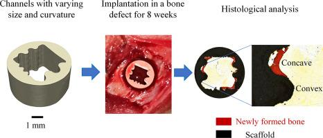
Bone has the capacity to regenerate itself for relatively small defects; however, this regenerative capacity is diminished in critical-size bone defects. The development of synthetic materials has risen as a distinct strategy to address this challenge. Effective synthetic materials to have emerged in recent years are bioceramic implants, which are biocompatible and highly bioactive. Yet nothing suitable for the repair of large bone defects has made the transition from laboratory to clinic. The clinical success of bioceramics has been shown to depend not only on the scaffold's intrinsic material properties but also on its internal porous geometry. This study aimed to systematically explore the implications of varying channel size, shape, and curvature in tissue scaffolds on in vivo bone regeneration outcomes. 3D printed bioceramic scaffolds with varying channel sizes (0.3 mm to 1.5 mm), shapes (circular vs rectangular), and curvatures (concave vs convex) were implanted in rabbit femoral defects for 8 weeks, followed by histological evaluation. We demonstrated that circular channel sizes of around 0.9 mm diameter significantly enhanced bone formation, compared to channel with diameters of 0.3 mm and 1.5 mm. Interestingly, varying channel shapes (rectangular vs circular) had no significant effect on the volume of newly formed bone. Furthermore, the present study systematically demonstrated the beneficial effect of concave surfaces on bone tissue growth in vivo, reinforcing previous in silico and in vitro findings. This study demonstrates that optimizing architectural configurations within ceramic scaffolds is crucial in enhancing bone regeneration outcomes.
Statement of significance
Despite the explosion of work on developing synthetic scaffolds to repair bone defects, the amount of new bone formed by scaffolds in vivo remains suboptimal. Recent studies have illuminated the pivotal role of scaffolds’ internal architecture in osteogenesis. However, these investigations have mostly remained confined to in silico and in vitro experiments. Among the in vivo studies conducted, there has been a lack of systematic analysis of individual architectural features. Herein, we utilized bioceramic 3D printing to conduct a systematic exploration of the effects of channel size, shape, and curvature on bone formation in vivo. Our results demonstrate the significant influence of channel size and curvature on in vivo outcomes. These findings provide invaluable insights into the design of more effective bone scaffolds.
期刊介绍:
Acta Biomaterialia is a monthly peer-reviewed scientific journal published by Elsevier. The journal was established in January 2005. The editor-in-chief is W.R. Wagner (University of Pittsburgh). The journal covers research in biomaterials science, including the interrelationship of biomaterial structure and function from macroscale to nanoscale. Topical coverage includes biomedical and biocompatible materials.


 分享
分享
 求助内容:
求助内容: 应助结果提醒方式:
 扫码关注我们
