Kathrin Becker, Henrike Ehrlich, Mira Hüfner, Nicole Rauch, Caroline Busch, Beryl Schwarz-Herzke, Dieter Drescher, Jürgen Becker
{"title":"新型 BW + 技术的资格以及不同成像方法在放射学龋齿检测中的灵敏度和特异性比较","authors":"Kathrin Becker, Henrike Ehrlich, Mira Hüfner, Nicole Rauch, Caroline Busch, Beryl Schwarz-Herzke, Dieter Drescher, Jürgen Becker","doi":"10.1007/s11282-024-00748-4","DOIUrl":null,"url":null,"abstract":"<h3 data-test=\"abstract-sub-heading\">Objectives</h3><p>Bitewing radiography is considered to be of high diagnostic value in caries detection, but owing to projections, lesions may remain undetected. The novel bitewing plus (BW +) technology enables scrolling through radiographs in different directions and angles. The present study aimed at comparing BW + with other 2D and 3D imaging methods in terms of sensitivity, specificity, and user reliability.</p><h3 data-test=\"abstract-sub-heading\">Materials and methods</h3><p>Five human cadavers were used in this study. In three cadavers, natural teeth were transplanted post-mortem. BW + , two-dimensional (digital sensors, imaging plates, 2D and 3D bitewing radiographs) and 3D methods (high and low dose CBCT) were taken. Carious lesions were evaluated on 96 teeth at three positions (mesial, distal, and occlusal) and scored according to their level of demineralization by ten observers, resulting in 35,799 possible lesions across all observers and settings. For reference, µCT scans of all teeth were performed.</p><h3 data-test=\"abstract-sub-heading\">Results</h3><p>Overall, radiographic evaluations showed a high rate of false-negative diagnoses, with around 70% of lesions remaining undetected, especially enamel lesions. BW + showed the highest sensitivity for dentinal caries and had comparatively high specificity overall.</p><h3 data-test=\"abstract-sub-heading\">Conclusions</h3><p>Within the limits of the study, BW + showed great potential for added diagnostic value, especially for dentinal caries. However, the tradeoff of diagnostic benefit and radiation exposure must be considered according to each patient’s age and risk.</p>","PeriodicalId":56103,"journal":{"name":"Oral Radiology","volume":"2017 1","pages":""},"PeriodicalIF":1.7000,"publicationDate":"2024-04-29","publicationTypes":"Journal Article","fieldsOfStudy":null,"isOpenAccess":false,"openAccessPdf":"","citationCount":"0","resultStr":"{\"title\":\"Eligibility of a novel BW + technology and comparison of sensitivity and specificity of different imaging methods for radiological caries detection\",\"authors\":\"Kathrin Becker, Henrike Ehrlich, Mira Hüfner, Nicole Rauch, Caroline Busch, Beryl Schwarz-Herzke, Dieter Drescher, Jürgen Becker\",\"doi\":\"10.1007/s11282-024-00748-4\",\"DOIUrl\":null,\"url\":null,\"abstract\":\"<h3 data-test=\\\"abstract-sub-heading\\\">Objectives</h3><p>Bitewing radiography is considered to be of high diagnostic value in caries detection, but owing to projections, lesions may remain undetected. The novel bitewing plus (BW +) technology enables scrolling through radiographs in different directions and angles. The present study aimed at comparing BW + with other 2D and 3D imaging methods in terms of sensitivity, specificity, and user reliability.</p><h3 data-test=\\\"abstract-sub-heading\\\">Materials and methods</h3><p>Five human cadavers were used in this study. In three cadavers, natural teeth were transplanted post-mortem. BW + , two-dimensional (digital sensors, imaging plates, 2D and 3D bitewing radiographs) and 3D methods (high and low dose CBCT) were taken. Carious lesions were evaluated on 96 teeth at three positions (mesial, distal, and occlusal) and scored according to their level of demineralization by ten observers, resulting in 35,799 possible lesions across all observers and settings. For reference, µCT scans of all teeth were performed.</p><h3 data-test=\\\"abstract-sub-heading\\\">Results</h3><p>Overall, radiographic evaluations showed a high rate of false-negative diagnoses, with around 70% of lesions remaining undetected, especially enamel lesions. BW + showed the highest sensitivity for dentinal caries and had comparatively high specificity overall.</p><h3 data-test=\\\"abstract-sub-heading\\\">Conclusions</h3><p>Within the limits of the study, BW + showed great potential for added diagnostic value, especially for dentinal caries. However, the tradeoff of diagnostic benefit and radiation exposure must be considered according to each patient’s age and risk.</p>\",\"PeriodicalId\":56103,\"journal\":{\"name\":\"Oral Radiology\",\"volume\":\"2017 1\",\"pages\":\"\"},\"PeriodicalIF\":1.7000,\"publicationDate\":\"2024-04-29\",\"publicationTypes\":\"Journal Article\",\"fieldsOfStudy\":null,\"isOpenAccess\":false,\"openAccessPdf\":\"\",\"citationCount\":\"0\",\"resultStr\":null,\"platform\":\"Semanticscholar\",\"paperid\":null,\"PeriodicalName\":\"Oral Radiology\",\"FirstCategoryId\":\"3\",\"ListUrlMain\":\"https://doi.org/10.1007/s11282-024-00748-4\",\"RegionNum\":3,\"RegionCategory\":\"医学\",\"ArticlePicture\":[],\"TitleCN\":null,\"AbstractTextCN\":null,\"PMCID\":null,\"EPubDate\":\"\",\"PubModel\":\"\",\"JCR\":\"Q3\",\"JCRName\":\"DENTISTRY, ORAL SURGERY & MEDICINE\",\"Score\":null,\"Total\":0}","platform":"Semanticscholar","paperid":null,"PeriodicalName":"Oral Radiology","FirstCategoryId":"3","ListUrlMain":"https://doi.org/10.1007/s11282-024-00748-4","RegionNum":3,"RegionCategory":"医学","ArticlePicture":[],"TitleCN":null,"AbstractTextCN":null,"PMCID":null,"EPubDate":"","PubModel":"","JCR":"Q3","JCRName":"DENTISTRY, ORAL SURGERY & MEDICINE","Score":null,"Total":0}
Eligibility of a novel BW + technology and comparison of sensitivity and specificity of different imaging methods for radiological caries detection
Objectives
Bitewing radiography is considered to be of high diagnostic value in caries detection, but owing to projections, lesions may remain undetected. The novel bitewing plus (BW +) technology enables scrolling through radiographs in different directions and angles. The present study aimed at comparing BW + with other 2D and 3D imaging methods in terms of sensitivity, specificity, and user reliability.
Materials and methods
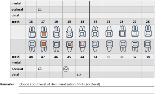
Five human cadavers were used in this study. In three cadavers, natural teeth were transplanted post-mortem. BW + , two-dimensional (digital sensors, imaging plates, 2D and 3D bitewing radiographs) and 3D methods (high and low dose CBCT) were taken. Carious lesions were evaluated on 96 teeth at three positions (mesial, distal, and occlusal) and scored according to their level of demineralization by ten observers, resulting in 35,799 possible lesions across all observers and settings. For reference, µCT scans of all teeth were performed.
Results
Overall, radiographic evaluations showed a high rate of false-negative diagnoses, with around 70% of lesions remaining undetected, especially enamel lesions. BW + showed the highest sensitivity for dentinal caries and had comparatively high specificity overall.
Conclusions
Within the limits of the study, BW + showed great potential for added diagnostic value, especially for dentinal caries. However, the tradeoff of diagnostic benefit and radiation exposure must be considered according to each patient’s age and risk.
期刊介绍:
As the official English-language journal of the Japanese Society for Oral and Maxillofacial Radiology and the Asian Academy of Oral and Maxillofacial Radiology, Oral Radiology is intended to be a forum for international collaboration in head and neck diagnostic imaging and all related fields. Oral Radiology features cutting-edge research papers, review articles, case reports, and technical notes from both the clinical and experimental fields. As membership in the Society is not a prerequisite, contributions are welcome from researchers and clinicians worldwide.


 分享
分享
 求助内容:
求助内容: 应助结果提醒方式:
 扫码关注我们
