Timothy S. Chisholm, Ronald Melki and Christopher A. Hunter*,
{"title":"配体轮廓分析作为一种诊断工具,用于区分患者衍生的α-突触核蛋白多态性","authors":"Timothy S. Chisholm, Ronald Melki and Christopher A. Hunter*, ","doi":"10.1021/acschemneuro.4c00178","DOIUrl":null,"url":null,"abstract":"<p >Amyloid fibrils are characteristic of many neurodegenerative diseases, including Alzheimer’s and Parkinson’s diseases. While different diseases may have fibrils formed of the same protein, the supramolecular morphology of these fibrils is disease-specific. Here, a method is reported to distinguish eight morphologically distinct amyloid fibrils based on differences in ligand binding properties. Eight fibrillar polymorphs of α-synuclein (αSyn) were investigated: five generated de novo using recombinant αSyn and three generated using protein misfolding cyclic amplification (PMCA) of recombinant αSyn seeded with brain homogenates from deceased patients diagnosed with Parkinson’s disease (PD), multiple system atrophy (MSA), and dementia with Lewy bodies (DLB). Fluorescence binding assays were carried out for each fibril using a toolkit of six different ligands. The fibril samples were separated into five categories based on a binary classification of whether they bound specific ligands or not. Quantitative binding measurements then allowed every fibrillar polymorph to be uniquely identified, and the PMCA fibrils derived from PD, MSA, and DLB patients could be unambiguously distinguished. This approach constitutes a novel and operationally simple method to differentiate amyloid fibril morphologies and to identify disease states using PMCA fibrils obtained by seeding with patient samples.</p>","PeriodicalId":13,"journal":{"name":"ACS Chemical Neuroscience","volume":"15 10","pages":"2080–2088"},"PeriodicalIF":4.5000,"publicationDate":"2024-05-01","publicationTypes":"Journal Article","fieldsOfStudy":null,"isOpenAccess":false,"openAccessPdf":"https://pubs.acs.org/doi/epdf/10.1021/acschemneuro.4c00178","citationCount":"0","resultStr":"{\"title\":\"Ligand Profiling as a Diagnostic Tool to Differentiate Patient-Derived α-Synuclein Polymorphs\",\"authors\":\"Timothy S. Chisholm, Ronald Melki and Christopher A. Hunter*, \",\"doi\":\"10.1021/acschemneuro.4c00178\",\"DOIUrl\":null,\"url\":null,\"abstract\":\"<p >Amyloid fibrils are characteristic of many neurodegenerative diseases, including Alzheimer’s and Parkinson’s diseases. While different diseases may have fibrils formed of the same protein, the supramolecular morphology of these fibrils is disease-specific. Here, a method is reported to distinguish eight morphologically distinct amyloid fibrils based on differences in ligand binding properties. Eight fibrillar polymorphs of α-synuclein (αSyn) were investigated: five generated de novo using recombinant αSyn and three generated using protein misfolding cyclic amplification (PMCA) of recombinant αSyn seeded with brain homogenates from deceased patients diagnosed with Parkinson’s disease (PD), multiple system atrophy (MSA), and dementia with Lewy bodies (DLB). Fluorescence binding assays were carried out for each fibril using a toolkit of six different ligands. The fibril samples were separated into five categories based on a binary classification of whether they bound specific ligands or not. Quantitative binding measurements then allowed every fibrillar polymorph to be uniquely identified, and the PMCA fibrils derived from PD, MSA, and DLB patients could be unambiguously distinguished. This approach constitutes a novel and operationally simple method to differentiate amyloid fibril morphologies and to identify disease states using PMCA fibrils obtained by seeding with patient samples.</p>\",\"PeriodicalId\":13,\"journal\":{\"name\":\"ACS Chemical Neuroscience\",\"volume\":\"15 10\",\"pages\":\"2080–2088\"},\"PeriodicalIF\":4.5000,\"publicationDate\":\"2024-05-01\",\"publicationTypes\":\"Journal Article\",\"fieldsOfStudy\":null,\"isOpenAccess\":false,\"openAccessPdf\":\"https://pubs.acs.org/doi/epdf/10.1021/acschemneuro.4c00178\",\"citationCount\":\"0\",\"resultStr\":null,\"platform\":\"Semanticscholar\",\"paperid\":null,\"PeriodicalName\":\"ACS Chemical Neuroscience\",\"FirstCategoryId\":\"3\",\"ListUrlMain\":\"https://pubs.acs.org/doi/10.1021/acschemneuro.4c00178\",\"RegionNum\":3,\"RegionCategory\":\"医学\",\"ArticlePicture\":[],\"TitleCN\":null,\"AbstractTextCN\":null,\"PMCID\":null,\"EPubDate\":\"\",\"PubModel\":\"\",\"JCR\":\"Q2\",\"JCRName\":\"BIOCHEMISTRY & MOLECULAR BIOLOGY\",\"Score\":null,\"Total\":0}","platform":"Semanticscholar","paperid":null,"PeriodicalName":"ACS Chemical Neuroscience","FirstCategoryId":"3","ListUrlMain":"https://pubs.acs.org/doi/10.1021/acschemneuro.4c00178","RegionNum":3,"RegionCategory":"医学","ArticlePicture":[],"TitleCN":null,"AbstractTextCN":null,"PMCID":null,"EPubDate":"","PubModel":"","JCR":"Q2","JCRName":"BIOCHEMISTRY & MOLECULAR BIOLOGY","Score":null,"Total":0}
Ligand Profiling as a Diagnostic Tool to Differentiate Patient-Derived α-Synuclein Polymorphs
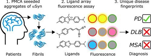
Amyloid fibrils are characteristic of many neurodegenerative diseases, including Alzheimer’s and Parkinson’s diseases. While different diseases may have fibrils formed of the same protein, the supramolecular morphology of these fibrils is disease-specific. Here, a method is reported to distinguish eight morphologically distinct amyloid fibrils based on differences in ligand binding properties. Eight fibrillar polymorphs of α-synuclein (αSyn) were investigated: five generated de novo using recombinant αSyn and three generated using protein misfolding cyclic amplification (PMCA) of recombinant αSyn seeded with brain homogenates from deceased patients diagnosed with Parkinson’s disease (PD), multiple system atrophy (MSA), and dementia with Lewy bodies (DLB). Fluorescence binding assays were carried out for each fibril using a toolkit of six different ligands. The fibril samples were separated into five categories based on a binary classification of whether they bound specific ligands or not. Quantitative binding measurements then allowed every fibrillar polymorph to be uniquely identified, and the PMCA fibrils derived from PD, MSA, and DLB patients could be unambiguously distinguished. This approach constitutes a novel and operationally simple method to differentiate amyloid fibril morphologies and to identify disease states using PMCA fibrils obtained by seeding with patient samples.
期刊介绍:
ACS Chemical Neuroscience publishes high-quality research articles and reviews that showcase chemical, quantitative biological, biophysical and bioengineering approaches to the understanding of the nervous system and to the development of new treatments for neurological disorders. Research in the journal focuses on aspects of chemical neurobiology and bio-neurochemistry such as the following:
Neurotransmitters and receptors
Neuropharmaceuticals and therapeutics
Neural development—Plasticity, and degeneration
Chemical, physical, and computational methods in neuroscience
Neuronal diseases—basis, detection, and treatment
Mechanism of aging, learning, memory and behavior
Pain and sensory processing
Neurotoxins
Neuroscience-inspired bioengineering
Development of methods in chemical neurobiology
Neuroimaging agents and technologies
Animal models for central nervous system diseases
Behavioral research


 分享
分享
 求助内容:
求助内容: 应助结果提醒方式:
 扫码关注我们
