{"title":"利用脲酶介导的混合 pH 指示剂的颜色混合,基于颜色演变的粘蛋白 1 检测灵敏传感器。","authors":"Jin-Hong Sui, Zhang-Run Xu","doi":"10.1016/j.talanta.2024.126191","DOIUrl":null,"url":null,"abstract":"<p><p>Mucin 1 is a significant tumor marker, and developing portable and cost-effective methods for its detection is crucial, especially in resource-limited areas. Herein, we developed an innovative approach for mucin 1 detection using a visible multicolor aptasensor. Urease-encapsulated DNA microspheres were used to mediate multicolor change facilitated by the color mixing of the mixed pH indicator, a mixed methyl red and bromocresol green solution. Distinct color changes were exhibited in response to varying mucin 1 concentrations. Notably, the color mixing of the mixed pH indicator was used to display various hues of colors, broadening the range of color variation. And color tonality is much easier to differentiate than color intensity, improving the resolution with naked-eyes. Besides, the variation of color from red to green (a pair of complementary colors) enhanced the color contrast, heightening sensitivity for visual detection. Importantly, the proposed method was successfully applied to detect mucin 1 in real samples, demonstrating a clear differentiation of colors between the samples of healthy individuals and breast cancer patients. The use of a mixed pH indicator as a multichromatic substrate offers the merits of low cost, fast response to pH variation, and plentiful color-evolution. And the incorporation of calcium carbonate microspheres to encapsulate urease ensures stable urease activity and avoids the need for extra urease decoration. The color-mixing dependent strategy opens a new way for multicolor detection of MUC1, characterized by vivid color changes.</p>","PeriodicalId":435,"journal":{"name":"Talanta","volume":"275 ","pages":"126191"},"PeriodicalIF":6.1000,"publicationDate":"2024-08-01","publicationTypes":"Journal Article","fieldsOfStudy":null,"isOpenAccess":false,"openAccessPdf":"","citationCount":"0","resultStr":"{\"title\":\"Profuse color-evolution based aptasensor for mucin 1 detection utilizing urease-mediated color mixing of the mixed pH indicator.\",\"authors\":\"Jin-Hong Sui, Zhang-Run Xu\",\"doi\":\"10.1016/j.talanta.2024.126191\",\"DOIUrl\":null,\"url\":null,\"abstract\":\"<p><p>Mucin 1 is a significant tumor marker, and developing portable and cost-effective methods for its detection is crucial, especially in resource-limited areas. Herein, we developed an innovative approach for mucin 1 detection using a visible multicolor aptasensor. Urease-encapsulated DNA microspheres were used to mediate multicolor change facilitated by the color mixing of the mixed pH indicator, a mixed methyl red and bromocresol green solution. Distinct color changes were exhibited in response to varying mucin 1 concentrations. Notably, the color mixing of the mixed pH indicator was used to display various hues of colors, broadening the range of color variation. And color tonality is much easier to differentiate than color intensity, improving the resolution with naked-eyes. Besides, the variation of color from red to green (a pair of complementary colors) enhanced the color contrast, heightening sensitivity for visual detection. Importantly, the proposed method was successfully applied to detect mucin 1 in real samples, demonstrating a clear differentiation of colors between the samples of healthy individuals and breast cancer patients. The use of a mixed pH indicator as a multichromatic substrate offers the merits of low cost, fast response to pH variation, and plentiful color-evolution. And the incorporation of calcium carbonate microspheres to encapsulate urease ensures stable urease activity and avoids the need for extra urease decoration. The color-mixing dependent strategy opens a new way for multicolor detection of MUC1, characterized by vivid color changes.</p>\",\"PeriodicalId\":435,\"journal\":{\"name\":\"Talanta\",\"volume\":\"275 \",\"pages\":\"126191\"},\"PeriodicalIF\":6.1000,\"publicationDate\":\"2024-08-01\",\"publicationTypes\":\"Journal Article\",\"fieldsOfStudy\":null,\"isOpenAccess\":false,\"openAccessPdf\":\"\",\"citationCount\":\"0\",\"resultStr\":null,\"platform\":\"Semanticscholar\",\"paperid\":null,\"PeriodicalName\":\"Talanta\",\"FirstCategoryId\":\"92\",\"ListUrlMain\":\"https://doi.org/10.1016/j.talanta.2024.126191\",\"RegionNum\":1,\"RegionCategory\":\"化学\",\"ArticlePicture\":[],\"TitleCN\":null,\"AbstractTextCN\":null,\"PMCID\":null,\"EPubDate\":\"2024/5/1 0:00:00\",\"PubModel\":\"Epub\",\"JCR\":\"Q1\",\"JCRName\":\"CHEMISTRY, ANALYTICAL\",\"Score\":null,\"Total\":0}","platform":"Semanticscholar","paperid":null,"PeriodicalName":"Talanta","FirstCategoryId":"92","ListUrlMain":"https://doi.org/10.1016/j.talanta.2024.126191","RegionNum":1,"RegionCategory":"化学","ArticlePicture":[],"TitleCN":null,"AbstractTextCN":null,"PMCID":null,"EPubDate":"2024/5/1 0:00:00","PubModel":"Epub","JCR":"Q1","JCRName":"CHEMISTRY, ANALYTICAL","Score":null,"Total":0}
Profuse color-evolution based aptasensor for mucin 1 detection utilizing urease-mediated color mixing of the mixed pH indicator.
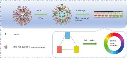
Mucin 1 is a significant tumor marker, and developing portable and cost-effective methods for its detection is crucial, especially in resource-limited areas. Herein, we developed an innovative approach for mucin 1 detection using a visible multicolor aptasensor. Urease-encapsulated DNA microspheres were used to mediate multicolor change facilitated by the color mixing of the mixed pH indicator, a mixed methyl red and bromocresol green solution. Distinct color changes were exhibited in response to varying mucin 1 concentrations. Notably, the color mixing of the mixed pH indicator was used to display various hues of colors, broadening the range of color variation. And color tonality is much easier to differentiate than color intensity, improving the resolution with naked-eyes. Besides, the variation of color from red to green (a pair of complementary colors) enhanced the color contrast, heightening sensitivity for visual detection. Importantly, the proposed method was successfully applied to detect mucin 1 in real samples, demonstrating a clear differentiation of colors between the samples of healthy individuals and breast cancer patients. The use of a mixed pH indicator as a multichromatic substrate offers the merits of low cost, fast response to pH variation, and plentiful color-evolution. And the incorporation of calcium carbonate microspheres to encapsulate urease ensures stable urease activity and avoids the need for extra urease decoration. The color-mixing dependent strategy opens a new way for multicolor detection of MUC1, characterized by vivid color changes.
期刊介绍:
Talanta provides a forum for the publication of original research papers, short communications, and critical reviews in all branches of pure and applied analytical chemistry. Papers are evaluated based on established guidelines, including the fundamental nature of the study, scientific novelty, substantial improvement or advantage over existing technology or methods, and demonstrated analytical applicability. Original research papers on fundamental studies, and on novel sensor and instrumentation developments, are encouraged. Novel or improved applications in areas such as clinical and biological chemistry, environmental analysis, geochemistry, materials science and engineering, and analytical platforms for omics development are welcome.
Analytical performance of methods should be determined, including interference and matrix effects, and methods should be validated by comparison with a standard method, or analysis of a certified reference material. Simple spiking recoveries may not be sufficient. The developed method should especially comprise information on selectivity, sensitivity, detection limits, accuracy, and reliability. However, applying official validation or robustness studies to a routine method or technique does not necessarily constitute novelty. Proper statistical treatment of the data should be provided. Relevant literature should be cited, including related publications by the authors, and authors should discuss how their proposed methodology compares with previously reported methods.


 分享
分享
 求助内容:
求助内容: 应助结果提醒方式:
 扫码关注我们
