{"title":"利用心脏磁共振成像技术对法布里心肌病和肥厚型心肌病进行分类的深度学习方法。","authors":"Wei-Wen Chen, Ling Kuo, Yi-Xun Lin, Wen-Chung Yu, Chien-Chao Tseng, Yenn-Jiang Lin, Ching-Chun Huang, Shih-Lin Chang, Jacky Chung-Hao Wu, Chun-Ku Chen, Ching-Yao Weng, Siwa Chan, Wei-Wen Lin, Yu-Cheng Hsieh, Ming-Chih Lin, Yun-Ching Fu, Tsung Chen, Shih-Ann Chen, Henry Horng-Shing Lu","doi":"10.1155/2024/6114826","DOIUrl":null,"url":null,"abstract":"<p><p>A challenge in accurately identifying and classifying left ventricular hypertrophy (LVH) is distinguishing it from hypertrophic cardiomyopathy (HCM) and Fabry disease. The reliance on imaging techniques often requires the expertise of multiple specialists, including cardiologists, radiologists, and geneticists. This variability in the interpretation and classification of LVH leads to inconsistent diagnoses. LVH, HCM, and Fabry cardiomyopathy can be differentiated using T1 mapping on cardiac magnetic resonance imaging (MRI). However, differentiation between HCM and Fabry cardiomyopathy using echocardiography or MRI cine images is challenging for cardiologists. Our proposed system named the MRI short-axis view left ventricular hypertrophy classifier (MSLVHC) is a high-accuracy standardized imaging classification model developed using AI and trained on MRI short-axis (SAX) view cine images to distinguish between HCM and Fabry disease. The model achieved impressive performance, with an <i>F</i>1-score of 0.846, an accuracy of 0.909, and an AUC of 0.914 when tested on the Taipei Veterans General Hospital (TVGH) dataset. Additionally, a single-blinding study and external testing using data from the Taichung Veterans General Hospital (TCVGH) demonstrated the reliability and effectiveness of the model, achieving an <i>F</i>1-score of 0.727, an accuracy of 0.806, and an AUC of 0.918, demonstrating the model's reliability and usefulness. This AI model holds promise as a valuable tool for assisting specialists in diagnosing LVH diseases.</p>","PeriodicalId":47063,"journal":{"name":"International Journal of Biomedical Imaging","volume":"2024 ","pages":"6114826"},"PeriodicalIF":1.3000,"publicationDate":"2024-04-26","publicationTypes":"Journal Article","fieldsOfStudy":null,"isOpenAccess":false,"openAccessPdf":"https://www.ncbi.nlm.nih.gov/pmc/articles/PMC11068448/pdf/","citationCount":"0","resultStr":"{\"title\":\"A Deep Learning Approach to Classify Fabry Cardiomyopathy from Hypertrophic Cardiomyopathy Using Cine Imaging on Cardiac Magnetic Resonance.\",\"authors\":\"Wei-Wen Chen, Ling Kuo, Yi-Xun Lin, Wen-Chung Yu, Chien-Chao Tseng, Yenn-Jiang Lin, Ching-Chun Huang, Shih-Lin Chang, Jacky Chung-Hao Wu, Chun-Ku Chen, Ching-Yao Weng, Siwa Chan, Wei-Wen Lin, Yu-Cheng Hsieh, Ming-Chih Lin, Yun-Ching Fu, Tsung Chen, Shih-Ann Chen, Henry Horng-Shing Lu\",\"doi\":\"10.1155/2024/6114826\",\"DOIUrl\":null,\"url\":null,\"abstract\":\"<p><p>A challenge in accurately identifying and classifying left ventricular hypertrophy (LVH) is distinguishing it from hypertrophic cardiomyopathy (HCM) and Fabry disease. The reliance on imaging techniques often requires the expertise of multiple specialists, including cardiologists, radiologists, and geneticists. This variability in the interpretation and classification of LVH leads to inconsistent diagnoses. LVH, HCM, and Fabry cardiomyopathy can be differentiated using T1 mapping on cardiac magnetic resonance imaging (MRI). However, differentiation between HCM and Fabry cardiomyopathy using echocardiography or MRI cine images is challenging for cardiologists. Our proposed system named the MRI short-axis view left ventricular hypertrophy classifier (MSLVHC) is a high-accuracy standardized imaging classification model developed using AI and trained on MRI short-axis (SAX) view cine images to distinguish between HCM and Fabry disease. The model achieved impressive performance, with an <i>F</i>1-score of 0.846, an accuracy of 0.909, and an AUC of 0.914 when tested on the Taipei Veterans General Hospital (TVGH) dataset. Additionally, a single-blinding study and external testing using data from the Taichung Veterans General Hospital (TCVGH) demonstrated the reliability and effectiveness of the model, achieving an <i>F</i>1-score of 0.727, an accuracy of 0.806, and an AUC of 0.918, demonstrating the model's reliability and usefulness. This AI model holds promise as a valuable tool for assisting specialists in diagnosing LVH diseases.</p>\",\"PeriodicalId\":47063,\"journal\":{\"name\":\"International Journal of Biomedical Imaging\",\"volume\":\"2024 \",\"pages\":\"6114826\"},\"PeriodicalIF\":1.3000,\"publicationDate\":\"2024-04-26\",\"publicationTypes\":\"Journal Article\",\"fieldsOfStudy\":null,\"isOpenAccess\":false,\"openAccessPdf\":\"https://www.ncbi.nlm.nih.gov/pmc/articles/PMC11068448/pdf/\",\"citationCount\":\"0\",\"resultStr\":null,\"platform\":\"Semanticscholar\",\"paperid\":null,\"PeriodicalName\":\"International Journal of Biomedical Imaging\",\"FirstCategoryId\":\"1085\",\"ListUrlMain\":\"https://doi.org/10.1155/2024/6114826\",\"RegionNum\":0,\"RegionCategory\":null,\"ArticlePicture\":[],\"TitleCN\":null,\"AbstractTextCN\":null,\"PMCID\":null,\"EPubDate\":\"2024/1/1 0:00:00\",\"PubModel\":\"eCollection\",\"JCR\":\"Q2\",\"JCRName\":\"ENGINEERING, BIOMEDICAL\",\"Score\":null,\"Total\":0}","platform":"Semanticscholar","paperid":null,"PeriodicalName":"International Journal of Biomedical Imaging","FirstCategoryId":"1085","ListUrlMain":"https://doi.org/10.1155/2024/6114826","RegionNum":0,"RegionCategory":null,"ArticlePicture":[],"TitleCN":null,"AbstractTextCN":null,"PMCID":null,"EPubDate":"2024/1/1 0:00:00","PubModel":"eCollection","JCR":"Q2","JCRName":"ENGINEERING, BIOMEDICAL","Score":null,"Total":0}
A Deep Learning Approach to Classify Fabry Cardiomyopathy from Hypertrophic Cardiomyopathy Using Cine Imaging on Cardiac Magnetic Resonance.
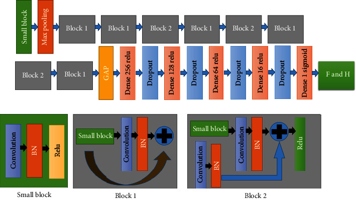
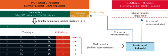
A challenge in accurately identifying and classifying left ventricular hypertrophy (LVH) is distinguishing it from hypertrophic cardiomyopathy (HCM) and Fabry disease. The reliance on imaging techniques often requires the expertise of multiple specialists, including cardiologists, radiologists, and geneticists. This variability in the interpretation and classification of LVH leads to inconsistent diagnoses. LVH, HCM, and Fabry cardiomyopathy can be differentiated using T1 mapping on cardiac magnetic resonance imaging (MRI). However, differentiation between HCM and Fabry cardiomyopathy using echocardiography or MRI cine images is challenging for cardiologists. Our proposed system named the MRI short-axis view left ventricular hypertrophy classifier (MSLVHC) is a high-accuracy standardized imaging classification model developed using AI and trained on MRI short-axis (SAX) view cine images to distinguish between HCM and Fabry disease. The model achieved impressive performance, with an F1-score of 0.846, an accuracy of 0.909, and an AUC of 0.914 when tested on the Taipei Veterans General Hospital (TVGH) dataset. Additionally, a single-blinding study and external testing using data from the Taichung Veterans General Hospital (TCVGH) demonstrated the reliability and effectiveness of the model, achieving an F1-score of 0.727, an accuracy of 0.806, and an AUC of 0.918, demonstrating the model's reliability and usefulness. This AI model holds promise as a valuable tool for assisting specialists in diagnosing LVH diseases.
期刊介绍:
The International Journal of Biomedical Imaging is managed by a board of editors comprising internationally renowned active researchers. The journal is freely accessible online and also offered for purchase in print format. It employs a web-based review system to ensure swift turnaround times while maintaining high standards. In addition to regular issues, special issues are organized by guest editors. The subject areas covered include (but are not limited to):
Digital radiography and tomosynthesis
X-ray computed tomography (CT)
Magnetic resonance imaging (MRI)
Single photon emission computed tomography (SPECT)
Positron emission tomography (PET)
Ultrasound imaging
Diffuse optical tomography, coherence, fluorescence, bioluminescence tomography, impedance tomography
Neutron imaging for biomedical applications
Magnetic and optical spectroscopy, and optical biopsy
Optical, electron, scanning tunneling/atomic force microscopy
Small animal imaging
Functional, cellular, and molecular imaging
Imaging assays for screening and molecular analysis
Microarray image analysis and bioinformatics
Emerging biomedical imaging techniques
Imaging modality fusion
Biomedical imaging instrumentation
Biomedical image processing, pattern recognition, and analysis
Biomedical image visualization, compression, transmission, and storage
Imaging and modeling related to systems biology and systems biomedicine
Applied mathematics, applied physics, and chemistry related to biomedical imaging
Grid-enabling technology for biomedical imaging and informatics


 分享
分享
 求助内容:
求助内容: 应助结果提醒方式:
 扫码关注我们
