Fang Bian, Fan-hong Niu, Ping-yuan Qu, Fang Gong, Jian-zhou Yan
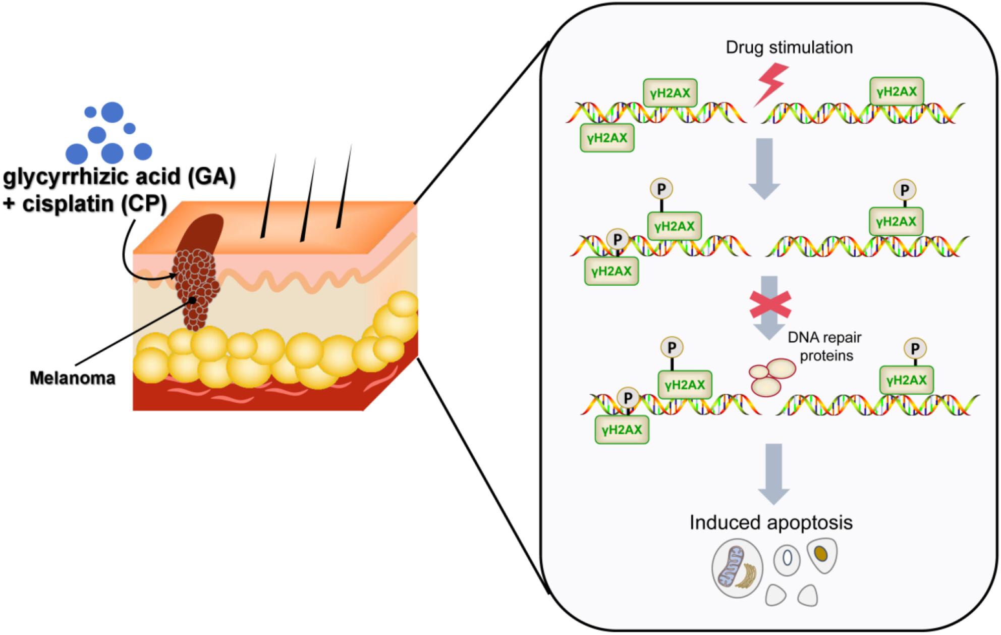
{"title":"甘草酸能抑制 DNA 损伤修复并增强顺铂诱导的黑色素瘤细胞凋亡。","authors":"Fang Bian, Fan-hong Niu, Ping-yuan Qu, Fang Gong, Jian-zhou Yan","doi":"10.1111/cbdd.14536","DOIUrl":null,"url":null,"abstract":"<p>This research was designed to prospect the mechanism and impact of glycyrrhizic acid (GA) on DNA damage repair and cisplatin (CP)-induced apoptosis of melanoma cells. First, human melanoma cell SK-MEL-28 was stimulated using GA for 24, 48, and 72 h. Then, the optimal treatment time and dosage were selected. After that, cell counting kit-8 (CCK-8) was employed for testing the cell viability, flow cytometry for the apoptosis, comet assay for the DNA damage of cells, and western blot for the cleaved-Caspase3, Caspase3, Bcl-2, and γH2AX protein expression levels. The experimental outcomes exhibited that as the GA concentration climbed up, the SK-MEL-28 cell viability dropped largely, while the apoptosis level raised significantly, especially at the concentration of 100 μm. In addition, compared with GA or CPtreatment only, CP combined with GA notably suppressed the viability of melanoma cells and promoted cell apoptosis at the cytological level. At the protein level, the combined treatment notably downregulated the Bcl-2 and Caspase3 expression levels, while significantly upregulated the cleaved-Caspase3 and γH2AX expression levels. Besides, CP + GA treatment promoted DNA damage at the DNA molecular level. Collectively, both GA and CP can inhibit DNA damage repair and enhance the apoptosis of SK-MEL-28 cells, and the synergistic treatment of both exhibits better efficacy.</p>","PeriodicalId":143,"journal":{"name":"Chemical Biology & Drug Design","volume":"103 5","pages":""},"PeriodicalIF":3.3000,"publicationDate":"2024-05-09","publicationTypes":"Journal Article","fieldsOfStudy":null,"isOpenAccess":false,"openAccessPdf":"","citationCount":"0","resultStr":"{\"title\":\"Glycyrrhizic acid inhibits DNA damage repair and enhances cisplatin-induced apoptosis of melanoma cells\",\"authors\":\"Fang Bian, Fan-hong Niu, Ping-yuan Qu, Fang Gong, Jian-zhou Yan\",\"doi\":\"10.1111/cbdd.14536\",\"DOIUrl\":null,\"url\":null,\"abstract\":\"<p>This research was designed to prospect the mechanism and impact of glycyrrhizic acid (GA) on DNA damage repair and cisplatin (CP)-induced apoptosis of melanoma cells. First, human melanoma cell SK-MEL-28 was stimulated using GA for 24, 48, and 72 h. Then, the optimal treatment time and dosage were selected. After that, cell counting kit-8 (CCK-8) was employed for testing the cell viability, flow cytometry for the apoptosis, comet assay for the DNA damage of cells, and western blot for the cleaved-Caspase3, Caspase3, Bcl-2, and γH2AX protein expression levels. The experimental outcomes exhibited that as the GA concentration climbed up, the SK-MEL-28 cell viability dropped largely, while the apoptosis level raised significantly, especially at the concentration of 100 μm. In addition, compared with GA or CPtreatment only, CP combined with GA notably suppressed the viability of melanoma cells and promoted cell apoptosis at the cytological level. At the protein level, the combined treatment notably downregulated the Bcl-2 and Caspase3 expression levels, while significantly upregulated the cleaved-Caspase3 and γH2AX expression levels. Besides, CP + GA treatment promoted DNA damage at the DNA molecular level. Collectively, both GA and CP can inhibit DNA damage repair and enhance the apoptosis of SK-MEL-28 cells, and the synergistic treatment of both exhibits better efficacy.</p>\",\"PeriodicalId\":143,\"journal\":{\"name\":\"Chemical Biology & Drug Design\",\"volume\":\"103 5\",\"pages\":\"\"},\"PeriodicalIF\":3.3000,\"publicationDate\":\"2024-05-09\",\"publicationTypes\":\"Journal Article\",\"fieldsOfStudy\":null,\"isOpenAccess\":false,\"openAccessPdf\":\"\",\"citationCount\":\"0\",\"resultStr\":null,\"platform\":\"Semanticscholar\",\"paperid\":null,\"PeriodicalName\":\"Chemical Biology & Drug Design\",\"FirstCategoryId\":\"3\",\"ListUrlMain\":\"https://onlinelibrary.wiley.com/doi/10.1111/cbdd.14536\",\"RegionNum\":4,\"RegionCategory\":\"医学\",\"ArticlePicture\":[],\"TitleCN\":null,\"AbstractTextCN\":null,\"PMCID\":null,\"EPubDate\":\"\",\"PubModel\":\"\",\"JCR\":\"Q2\",\"JCRName\":\"BIOCHEMISTRY & MOLECULAR BIOLOGY\",\"Score\":null,\"Total\":0}","platform":"Semanticscholar","paperid":null,"PeriodicalName":"Chemical Biology & Drug Design","FirstCategoryId":"3","ListUrlMain":"https://onlinelibrary.wiley.com/doi/10.1111/cbdd.14536","RegionNum":4,"RegionCategory":"医学","ArticlePicture":[],"TitleCN":null,"AbstractTextCN":null,"PMCID":null,"EPubDate":"","PubModel":"","JCR":"Q2","JCRName":"BIOCHEMISTRY & MOLECULAR BIOLOGY","Score":null,"Total":0}
引用次数: 0
摘要
本研究旨在探讨甘草酸(GA)对DNA损伤修复和顺铂(CP)诱导黑色素瘤细胞凋亡的机制和影响。首先,使用甘草酸刺激人黑色素瘤细胞SK-MEL-28 24、48和72小时,然后选择最佳治疗时间和剂量。然后,采用细胞计数试剂盒-8(CCK-8)检测细胞活力,流式细胞仪检测细胞凋亡,彗星试验检测细胞的 DNA 损伤,Western 印迹检测裂解-Caspase3、Caspase3、Bcl-2 和 γH2AX 蛋白表达水平。实验结果表明,随着 GA 浓度的升高,SK-MEL-28 细胞的存活率大幅下降,而细胞凋亡水平则显著上升,尤其是在浓度为 100 μm 时。此外,与只用 GA 或 CP 处理相比,CP 与 GA 联合处理在细胞学水平上明显抑制了黑色素瘤细胞的活力,促进了细胞凋亡。在蛋白质水平上,联合处理显著下调了Bcl-2和Caspase3的表达水平,同时显著上调了裂解的Caspase3和γH2AX的表达水平。此外,CP + GA 处理在 DNA 分子水平上促进了 DNA 损伤。综上所述,GA和CP均可抑制DNA损伤修复,促进SK-MEL-28细胞凋亡,二者协同处理效果更佳。
Glycyrrhizic acid inhibits DNA damage repair and enhances cisplatin-induced apoptosis of melanoma cells
This research was designed to prospect the mechanism and impact of glycyrrhizic acid (GA) on DNA damage repair and cisplatin (CP)-induced apoptosis of melanoma cells. First, human melanoma cell SK-MEL-28 was stimulated using GA for 24, 48, and 72 h. Then, the optimal treatment time and dosage were selected. After that, cell counting kit-8 (CCK-8) was employed for testing the cell viability, flow cytometry for the apoptosis, comet assay for the DNA damage of cells, and western blot for the cleaved-Caspase3, Caspase3, Bcl-2, and γH2AX protein expression levels. The experimental outcomes exhibited that as the GA concentration climbed up, the SK-MEL-28 cell viability dropped largely, while the apoptosis level raised significantly, especially at the concentration of 100 μm. In addition, compared with GA or CPtreatment only, CP combined with GA notably suppressed the viability of melanoma cells and promoted cell apoptosis at the cytological level. At the protein level, the combined treatment notably downregulated the Bcl-2 and Caspase3 expression levels, while significantly upregulated the cleaved-Caspase3 and γH2AX expression levels. Besides, CP + GA treatment promoted DNA damage at the DNA molecular level. Collectively, both GA and CP can inhibit DNA damage repair and enhance the apoptosis of SK-MEL-28 cells, and the synergistic treatment of both exhibits better efficacy.
期刊介绍:
Chemical Biology & Drug Design is a peer-reviewed scientific journal that is dedicated to the advancement of innovative science, technology and medicine with a focus on the multidisciplinary fields of chemical biology and drug design. It is the aim of Chemical Biology & Drug Design to capture significant research and drug discovery that highlights new concepts, insight and new findings within the scope of chemical biology and drug design.


 分享
分享
 求助内容:
求助内容: 应助结果提醒方式:
 扫码关注我们
