{"title":"通过心脏计算机断层扫描评估冠状动脉的高细胞外容积分数患者的临床特征。","authors":"Tetsuya Oguni, Seiji Takashio, Naoto Kuyama, Kyoko Hirakawa, Shinsuke Hanatani, Fumi Oike, Hiroki Usuku, Yasushi Matsuzawa, Masafumi Kidoh, Seitaro Oda, Eiichiro Yamamoto, Mitsuharu Ueda, Toshinori Hirai, Kenichi Tsujita","doi":"10.1093/ehjopen/oeae036","DOIUrl":null,"url":null,"abstract":"<p><strong>Aims: </strong>This study aims to evaluate the distribution of extracellular volume fraction detected via computed tomography, clinical characteristics of high extracellular volume fraction detected via computed tomography, and the rate of incidental detection of cardiac amyloidosis in patients undergoing cardiac computed tomography for coronary artery evaluation.</p><p><strong>Methods and results: </strong>This study included 874 consecutive patients (mean age, 74.4 ± 7.1 years; men, 65%), comprising men aged ≥60 years and women aged ≥70 years, who had undergone cardiac computed tomography between January 2020 and September 2022. The mean extracellular volume fraction detected via computed tomography was 29.7 ± 5.2%, and 108 patients (12.4%) had an extracellular volume fraction detected via computed tomography of ≥35%. Older age (75.9 ± 8.2 years vs. 74.2 ± 6.9 years; <i>P</i> = 0.042), male sex (75.9% vs. 63.0%; <i>P</i> = 0.007), impaired left ventricular ejection fraction, increased high-sensitivity cardiac troponin T and B-type natriuretic peptide levels, and increased left ventricular thickness showed significant associations with an extracellular volume fraction detected via computed tomography of ≥35%. Cardiac amyloidosis was diagnosed incidentally in 15 patients based on an increase in extracellular volume fraction detected via computed tomography. The prevalence of cardiac amyloidosis was 1.7% (15/874) and 14.3% (15/105) in the entire study population and in patients with an extracellular volume fraction detected via computed tomography of ≥35%, respectively. An increase in the extracellular volume fraction detected via computed tomography was suggestive of cardiac amyloidosis.</p><p><strong>Conclusion: </strong>Elevated extracellular volume fraction detected via computed tomography, associated with elevated cardiac biomarker levels and myocardial structural changes, may lead to the incidental diagnosis of cardiac amyloidosis.</p>","PeriodicalId":93995,"journal":{"name":"European heart journal open","volume":"4 3","pages":"oeae036"},"PeriodicalIF":0.0000,"publicationDate":"2024-04-27","publicationTypes":"Journal Article","fieldsOfStudy":null,"isOpenAccess":false,"openAccessPdf":"https://www.ncbi.nlm.nih.gov/pmc/articles/PMC11095558/pdf/","citationCount":"0","resultStr":"{\"title\":\"Clinical characteristics of patients with high extracellular volume fraction evaluated by cardiac computed tomography for coronary artery evaluation.\",\"authors\":\"Tetsuya Oguni, Seiji Takashio, Naoto Kuyama, Kyoko Hirakawa, Shinsuke Hanatani, Fumi Oike, Hiroki Usuku, Yasushi Matsuzawa, Masafumi Kidoh, Seitaro Oda, Eiichiro Yamamoto, Mitsuharu Ueda, Toshinori Hirai, Kenichi Tsujita\",\"doi\":\"10.1093/ehjopen/oeae036\",\"DOIUrl\":null,\"url\":null,\"abstract\":\"<p><strong>Aims: </strong>This study aims to evaluate the distribution of extracellular volume fraction detected via computed tomography, clinical characteristics of high extracellular volume fraction detected via computed tomography, and the rate of incidental detection of cardiac amyloidosis in patients undergoing cardiac computed tomography for coronary artery evaluation.</p><p><strong>Methods and results: </strong>This study included 874 consecutive patients (mean age, 74.4 ± 7.1 years; men, 65%), comprising men aged ≥60 years and women aged ≥70 years, who had undergone cardiac computed tomography between January 2020 and September 2022. The mean extracellular volume fraction detected via computed tomography was 29.7 ± 5.2%, and 108 patients (12.4%) had an extracellular volume fraction detected via computed tomography of ≥35%. Older age (75.9 ± 8.2 years vs. 74.2 ± 6.9 years; <i>P</i> = 0.042), male sex (75.9% vs. 63.0%; <i>P</i> = 0.007), impaired left ventricular ejection fraction, increased high-sensitivity cardiac troponin T and B-type natriuretic peptide levels, and increased left ventricular thickness showed significant associations with an extracellular volume fraction detected via computed tomography of ≥35%. Cardiac amyloidosis was diagnosed incidentally in 15 patients based on an increase in extracellular volume fraction detected via computed tomography. The prevalence of cardiac amyloidosis was 1.7% (15/874) and 14.3% (15/105) in the entire study population and in patients with an extracellular volume fraction detected via computed tomography of ≥35%, respectively. An increase in the extracellular volume fraction detected via computed tomography was suggestive of cardiac amyloidosis.</p><p><strong>Conclusion: </strong>Elevated extracellular volume fraction detected via computed tomography, associated with elevated cardiac biomarker levels and myocardial structural changes, may lead to the incidental diagnosis of cardiac amyloidosis.</p>\",\"PeriodicalId\":93995,\"journal\":{\"name\":\"European heart journal open\",\"volume\":\"4 3\",\"pages\":\"oeae036\"},\"PeriodicalIF\":0.0000,\"publicationDate\":\"2024-04-27\",\"publicationTypes\":\"Journal Article\",\"fieldsOfStudy\":null,\"isOpenAccess\":false,\"openAccessPdf\":\"https://www.ncbi.nlm.nih.gov/pmc/articles/PMC11095558/pdf/\",\"citationCount\":\"0\",\"resultStr\":null,\"platform\":\"Semanticscholar\",\"paperid\":null,\"PeriodicalName\":\"European heart journal open\",\"FirstCategoryId\":\"1085\",\"ListUrlMain\":\"https://doi.org/10.1093/ehjopen/oeae036\",\"RegionNum\":0,\"RegionCategory\":null,\"ArticlePicture\":[],\"TitleCN\":null,\"AbstractTextCN\":null,\"PMCID\":null,\"EPubDate\":\"2024/5/1 0:00:00\",\"PubModel\":\"eCollection\",\"JCR\":\"\",\"JCRName\":\"\",\"Score\":null,\"Total\":0}","platform":"Semanticscholar","paperid":null,"PeriodicalName":"European heart journal open","FirstCategoryId":"1085","ListUrlMain":"https://doi.org/10.1093/ehjopen/oeae036","RegionNum":0,"RegionCategory":null,"ArticlePicture":[],"TitleCN":null,"AbstractTextCN":null,"PMCID":null,"EPubDate":"2024/5/1 0:00:00","PubModel":"eCollection","JCR":"","JCRName":"","Score":null,"Total":0}
Clinical characteristics of patients with high extracellular volume fraction evaluated by cardiac computed tomography for coronary artery evaluation.
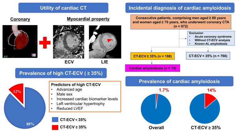
Aims: This study aims to evaluate the distribution of extracellular volume fraction detected via computed tomography, clinical characteristics of high extracellular volume fraction detected via computed tomography, and the rate of incidental detection of cardiac amyloidosis in patients undergoing cardiac computed tomography for coronary artery evaluation.
Methods and results: This study included 874 consecutive patients (mean age, 74.4 ± 7.1 years; men, 65%), comprising men aged ≥60 years and women aged ≥70 years, who had undergone cardiac computed tomography between January 2020 and September 2022. The mean extracellular volume fraction detected via computed tomography was 29.7 ± 5.2%, and 108 patients (12.4%) had an extracellular volume fraction detected via computed tomography of ≥35%. Older age (75.9 ± 8.2 years vs. 74.2 ± 6.9 years; P = 0.042), male sex (75.9% vs. 63.0%; P = 0.007), impaired left ventricular ejection fraction, increased high-sensitivity cardiac troponin T and B-type natriuretic peptide levels, and increased left ventricular thickness showed significant associations with an extracellular volume fraction detected via computed tomography of ≥35%. Cardiac amyloidosis was diagnosed incidentally in 15 patients based on an increase in extracellular volume fraction detected via computed tomography. The prevalence of cardiac amyloidosis was 1.7% (15/874) and 14.3% (15/105) in the entire study population and in patients with an extracellular volume fraction detected via computed tomography of ≥35%, respectively. An increase in the extracellular volume fraction detected via computed tomography was suggestive of cardiac amyloidosis.
Conclusion: Elevated extracellular volume fraction detected via computed tomography, associated with elevated cardiac biomarker levels and myocardial structural changes, may lead to the incidental diagnosis of cardiac amyloidosis.


 分享
分享

 求助内容:
求助内容: 应助结果提醒方式:
 扫码关注我们
